Blog
Credibly reintermediate backend ideas for cross-platform models. Continually reintermediate integrated processes through technically sound intellectual capital.
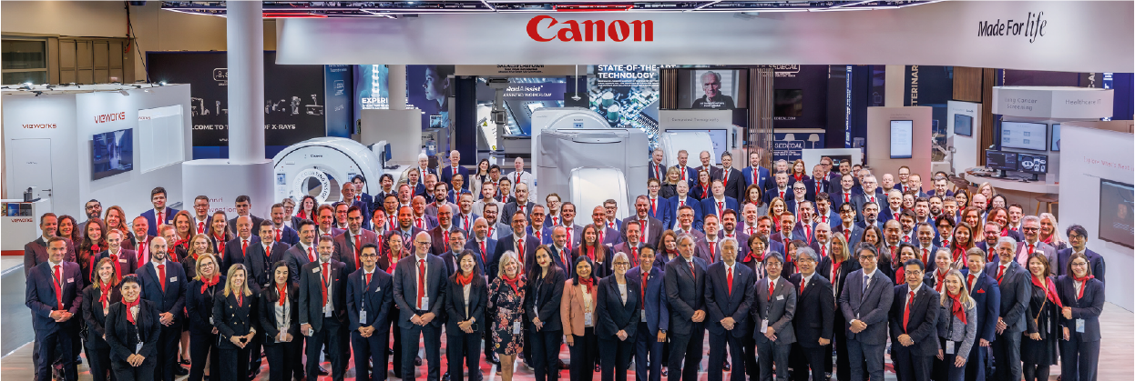
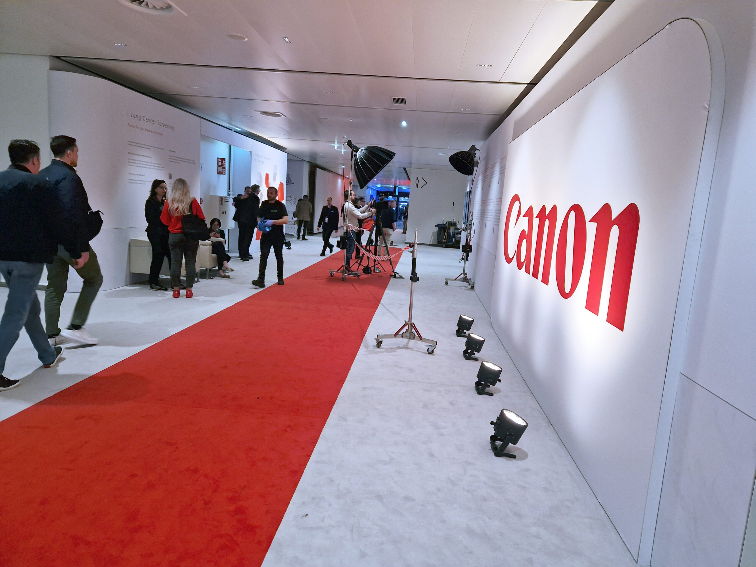
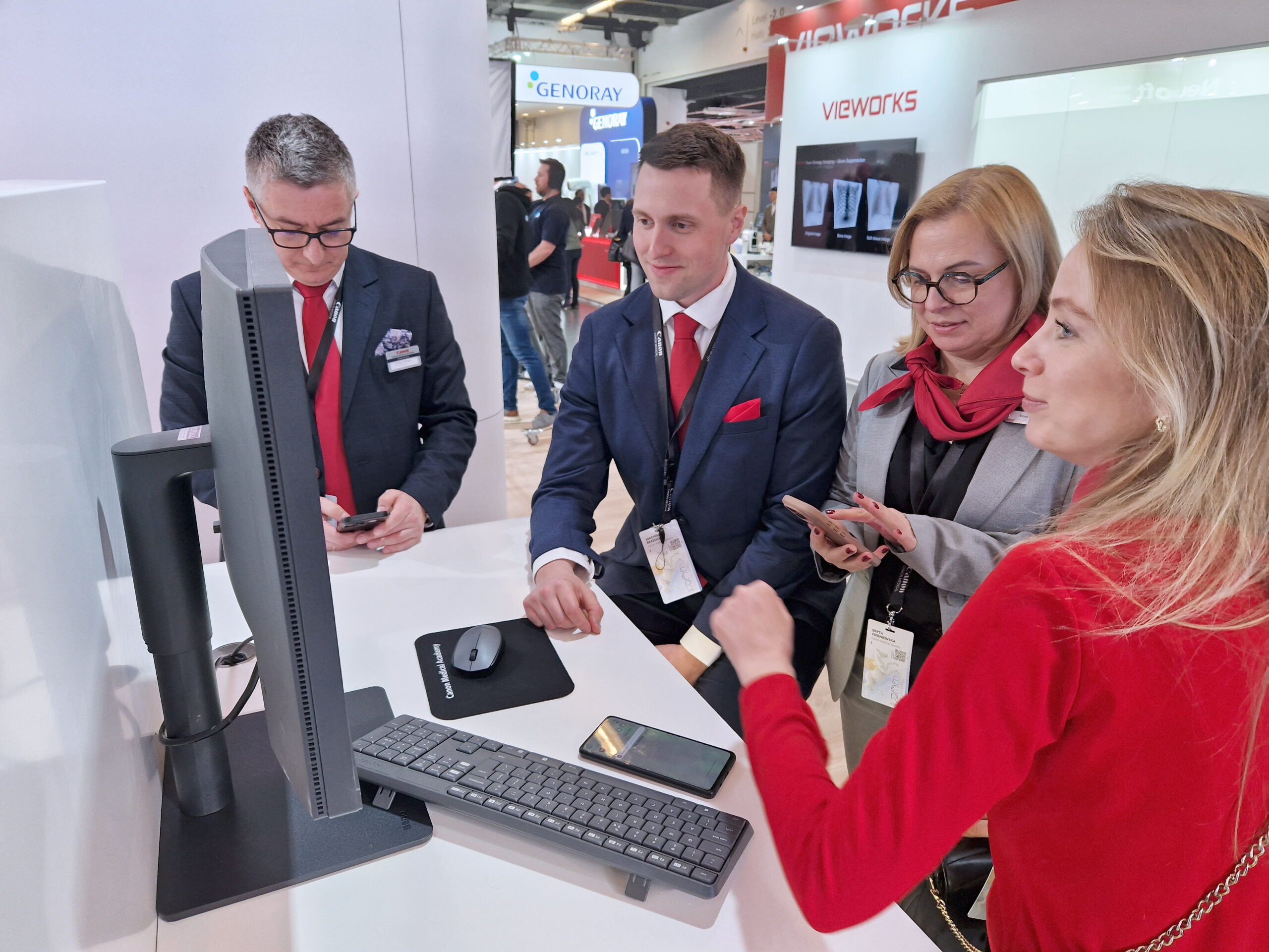
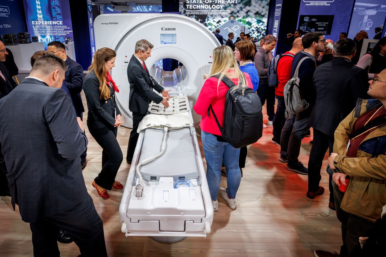
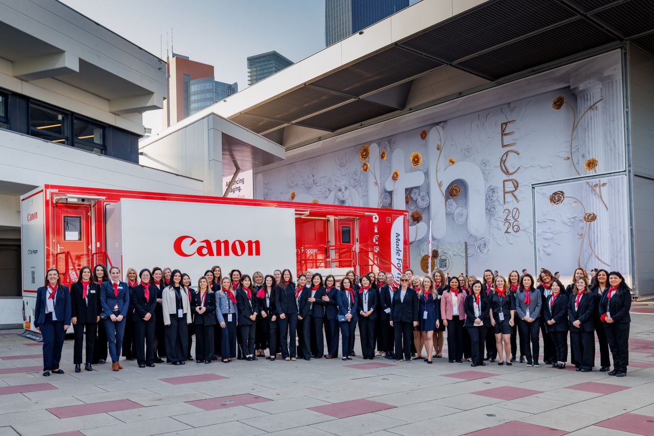
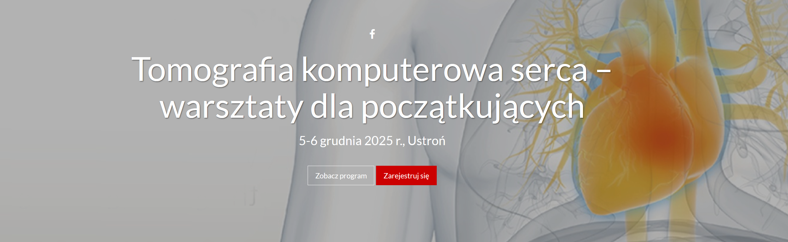
ECR 2026 | Satellite Symposium | Canon Medical Academy
Advancing CEUS in Radiology: Clinical Innovations, Faster Pathways, Higher Efficiency | Symposium
Thursday, March 5, 2026, 14:30–15:30 (60 min) | Room G2
ECR 2026 | Satellite Symposium | Canon Medical Academy
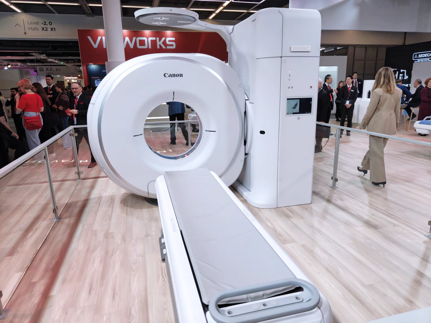
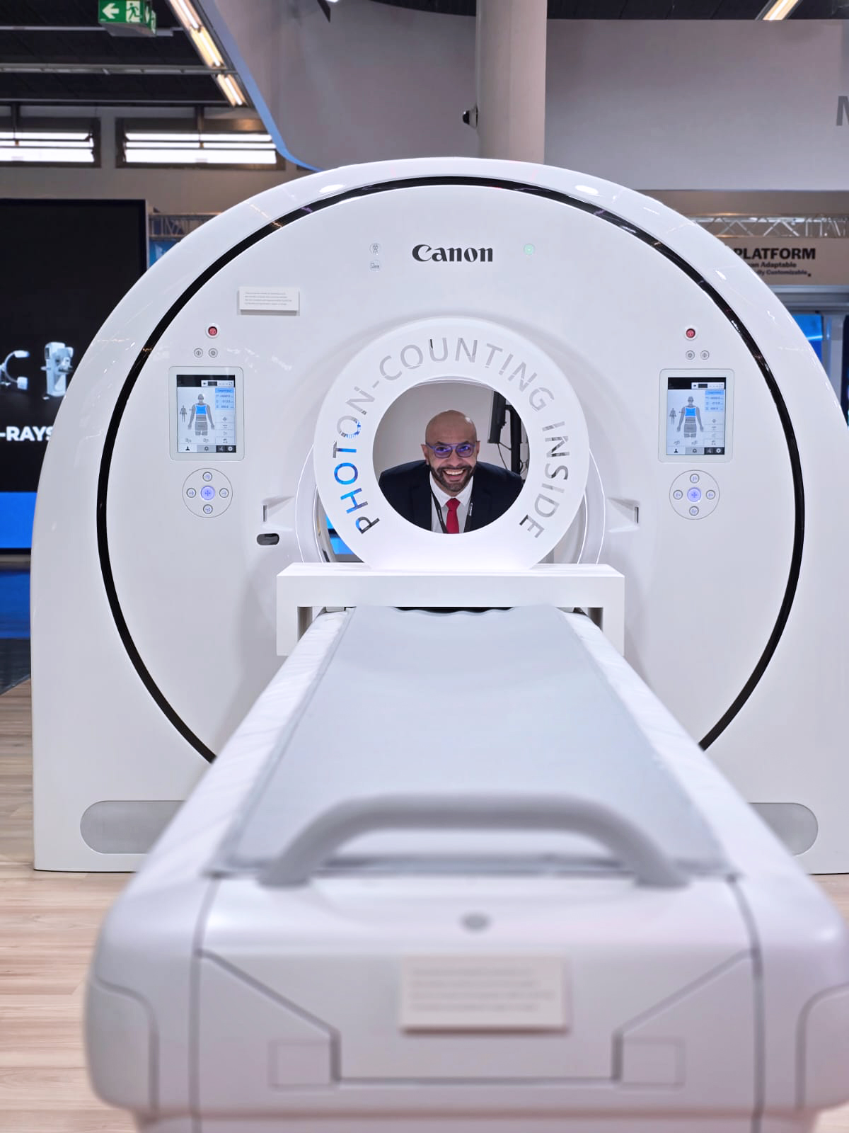
The Next Level of Diagnostic Quality in Photon Counting and Area Detector CT: AI, Spectral, and UHR | Symposium
Friday, March 6, 2026, 09:30–10:30 (60min), Room N
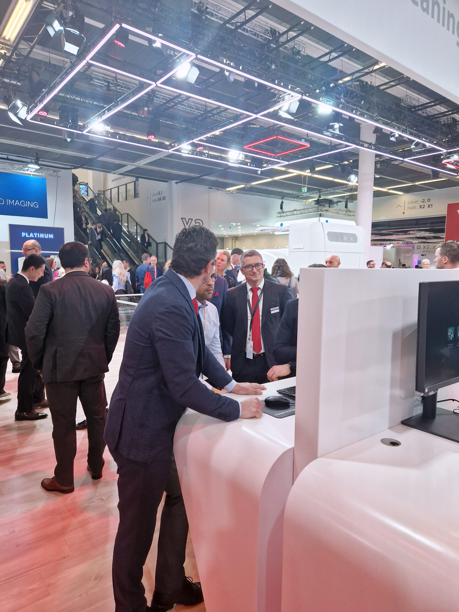
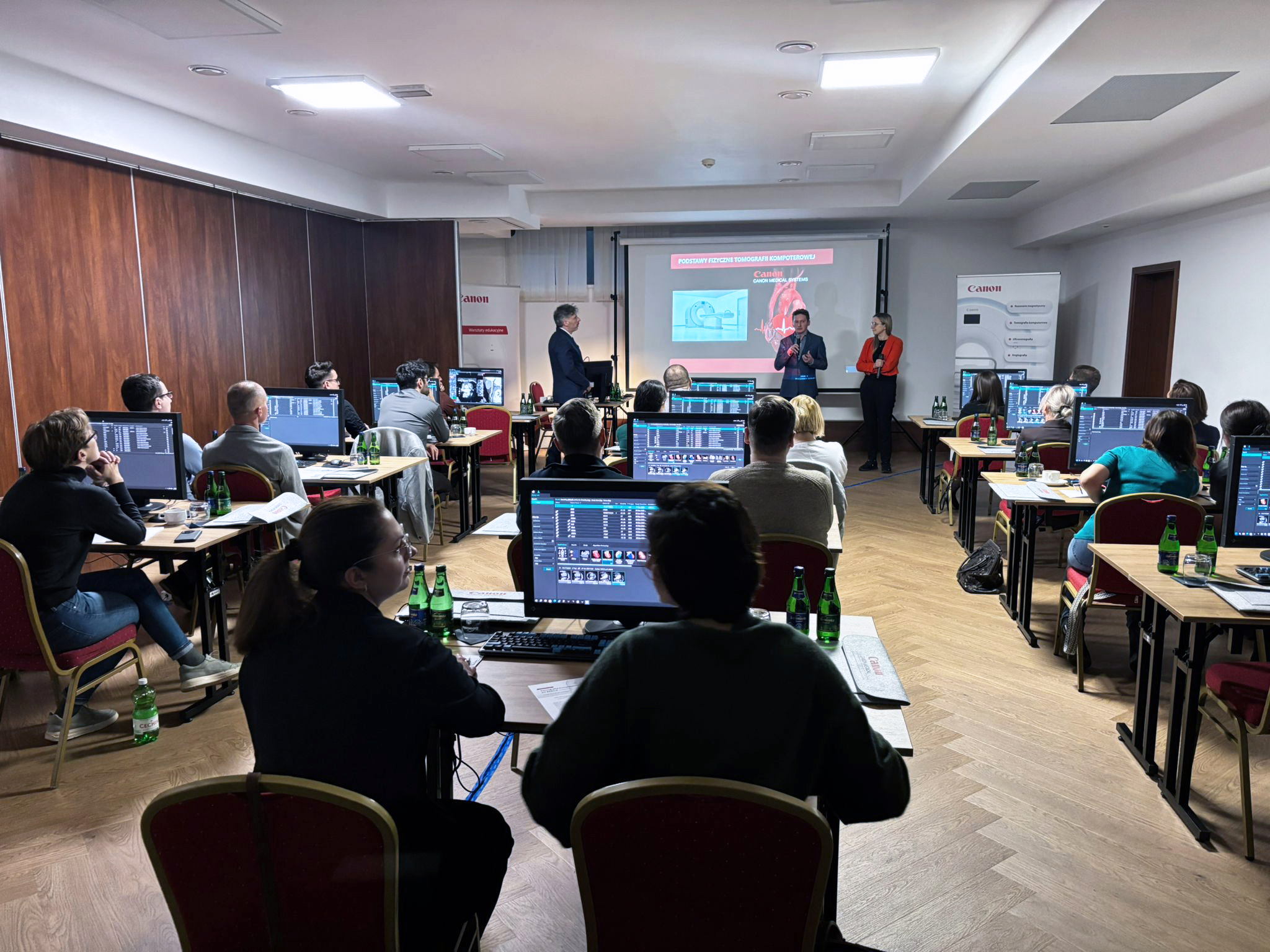
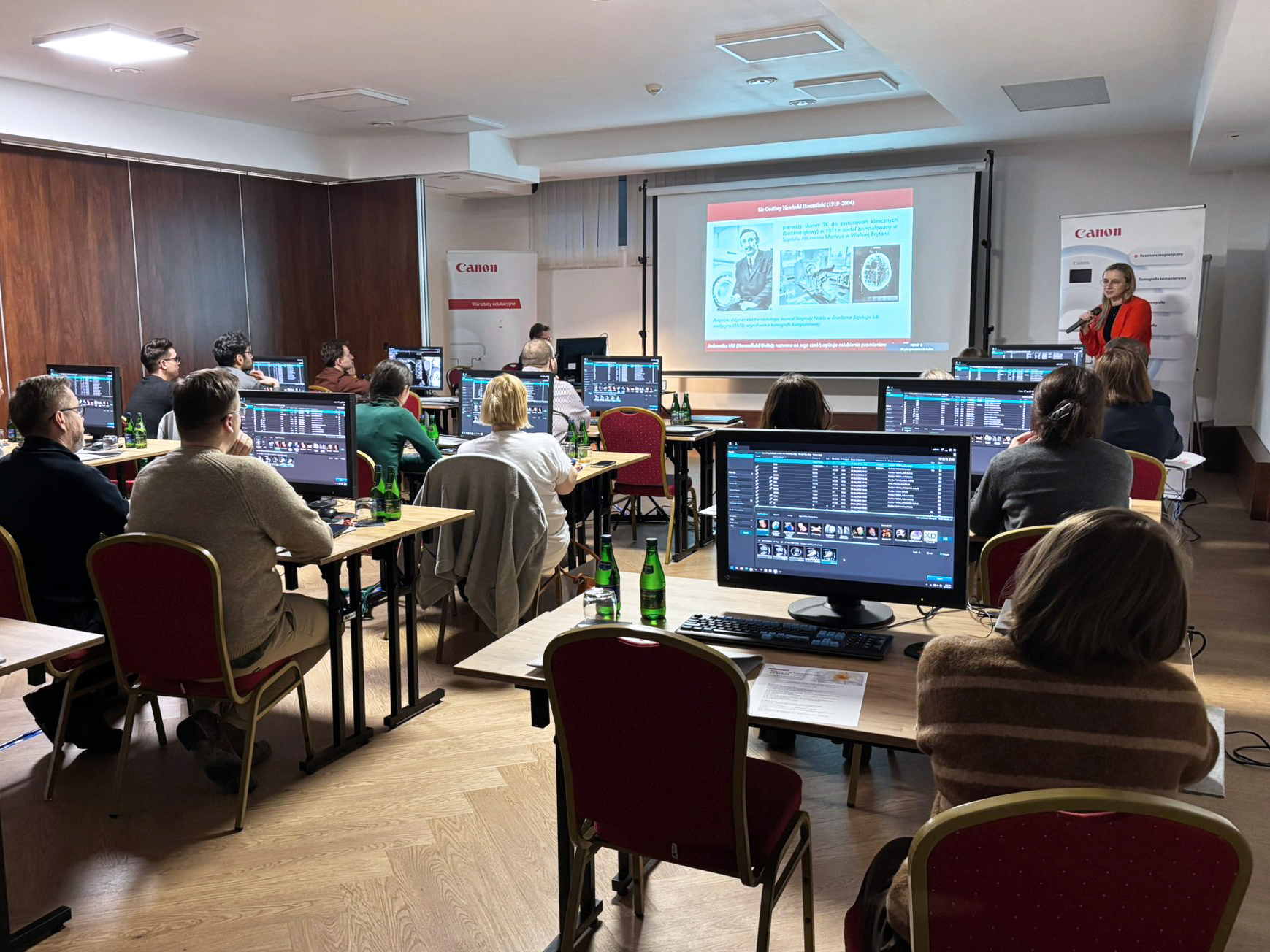
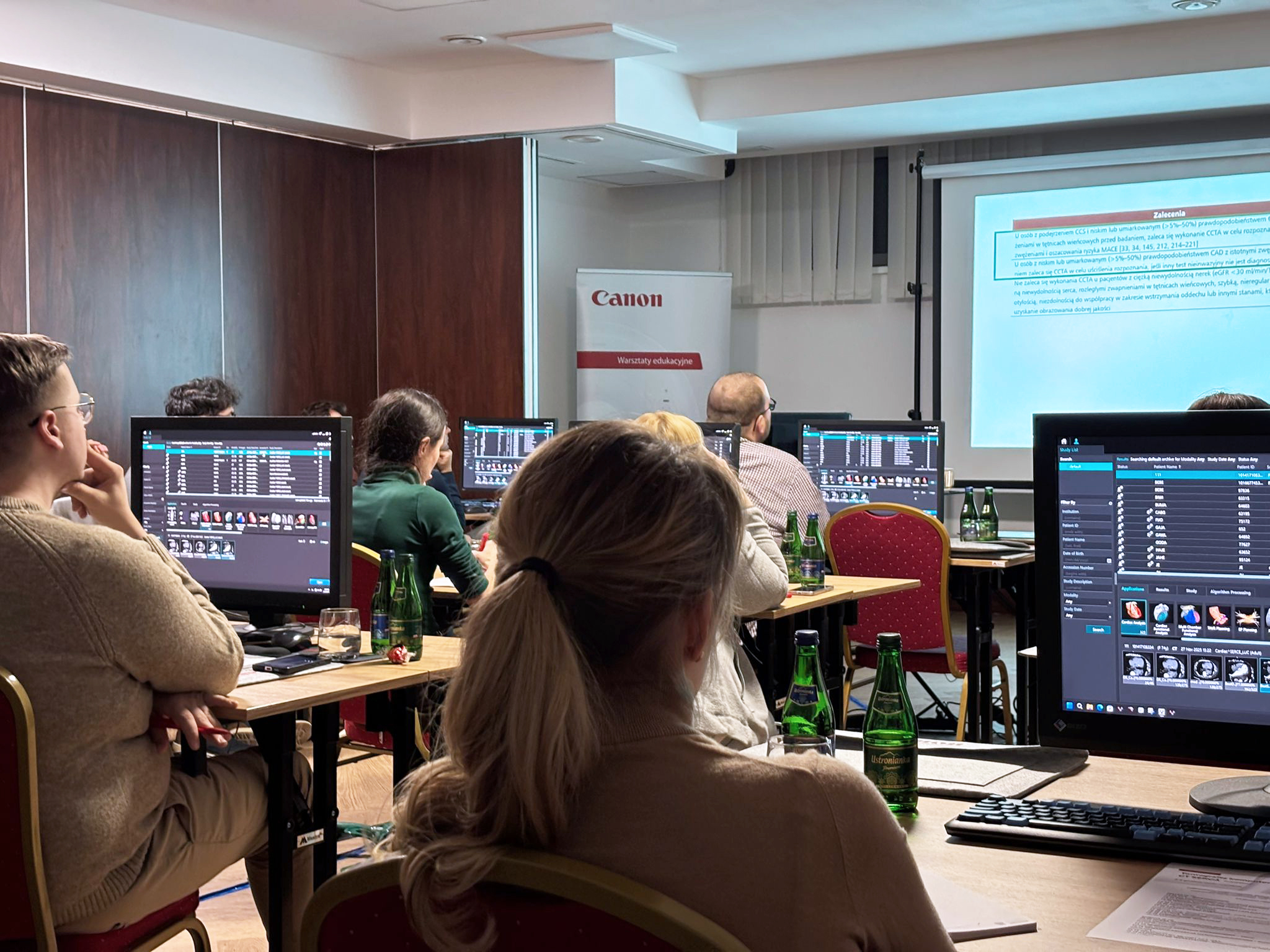
ECR 2026 | WORKSHOPS | Canon Medical Academy
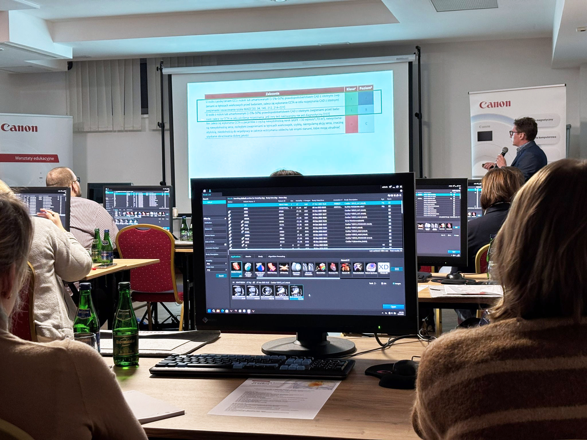
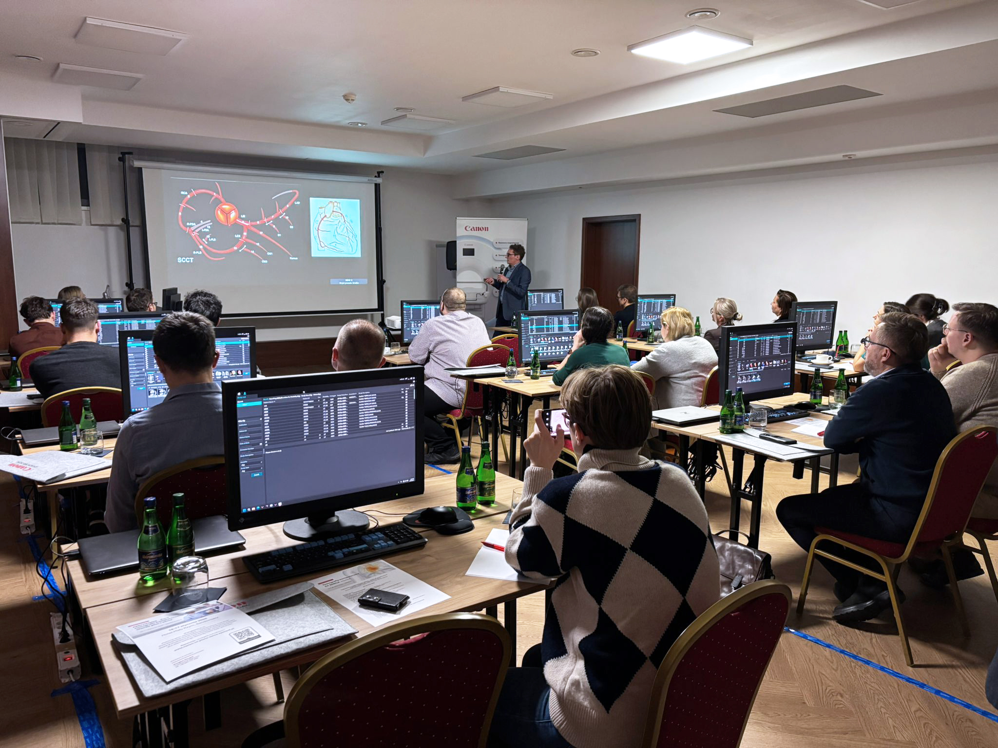
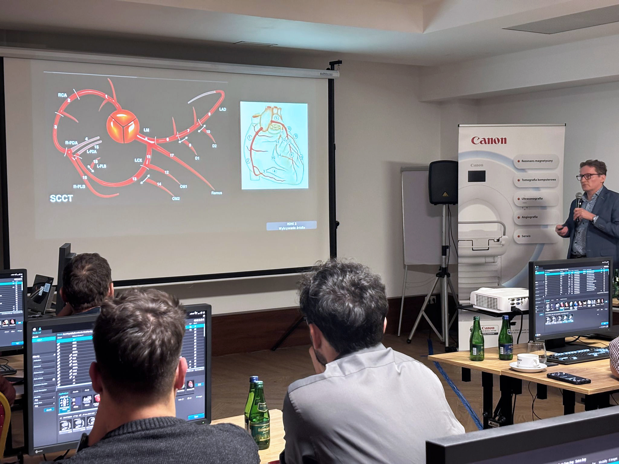
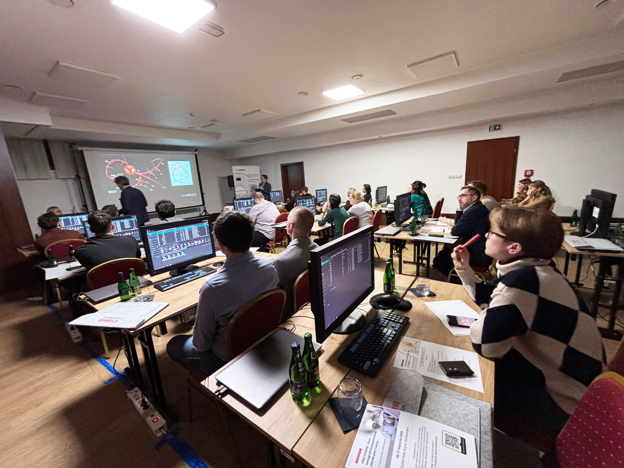
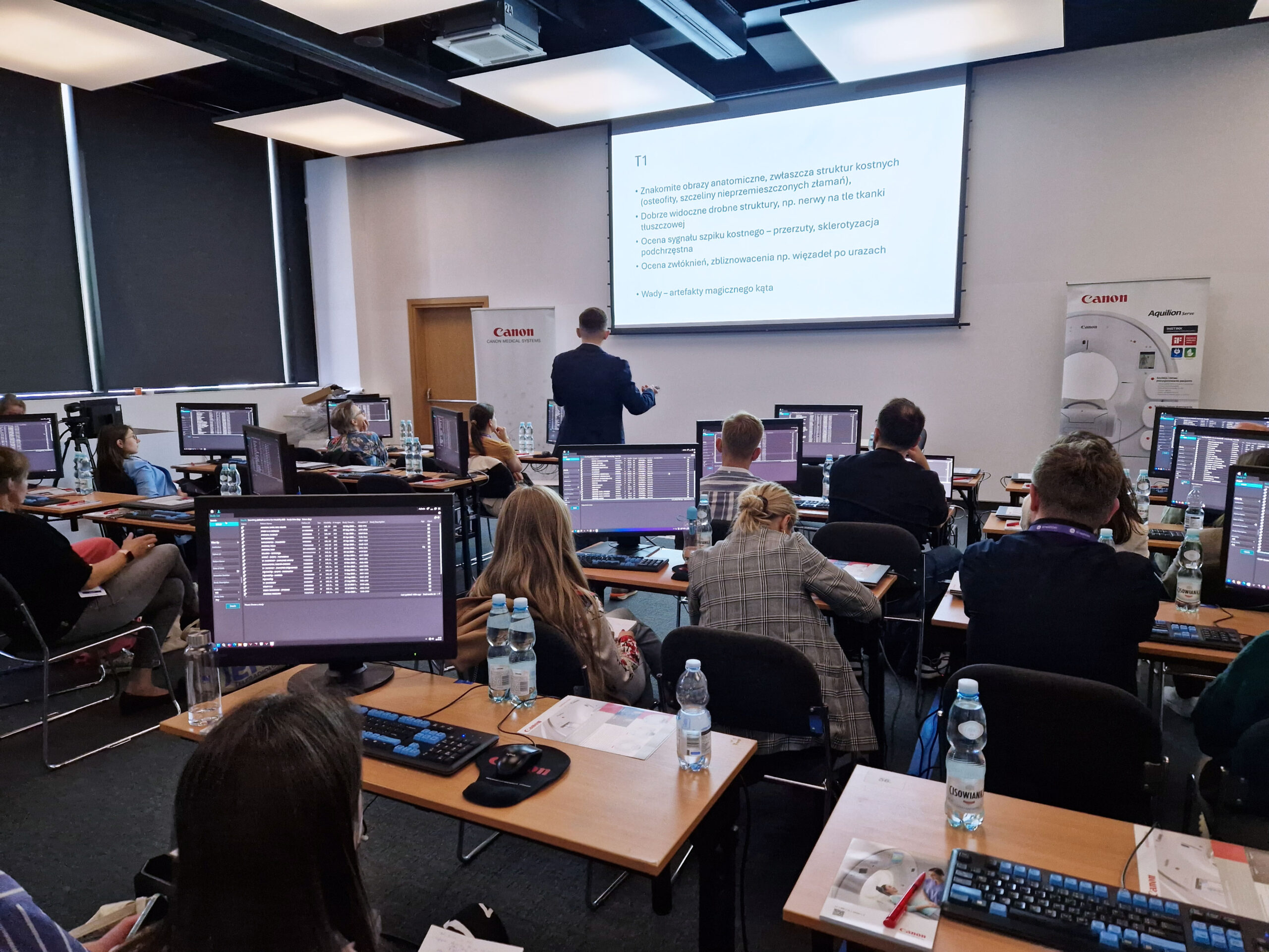
Workshops provided by Canon Medical Academy
Wednesday, March 4
Thursday, March 5
Friday, March 6
Saturday, March 7
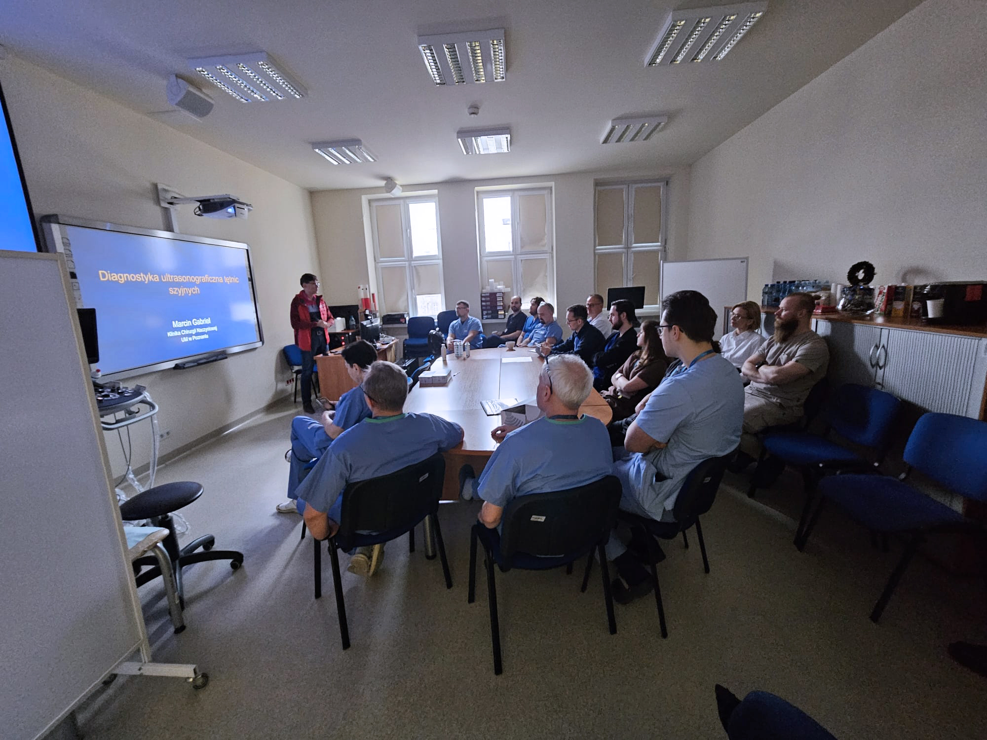
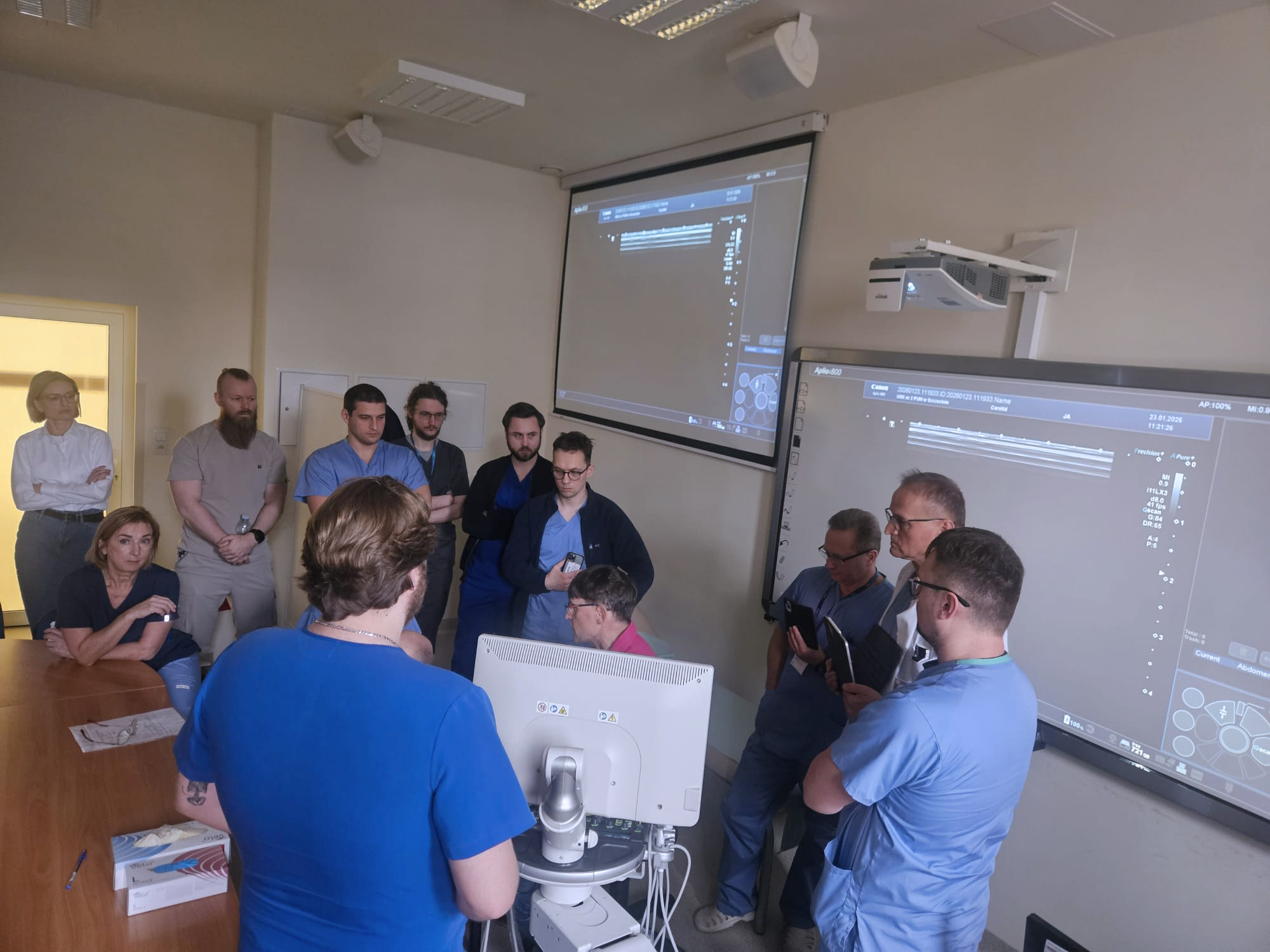
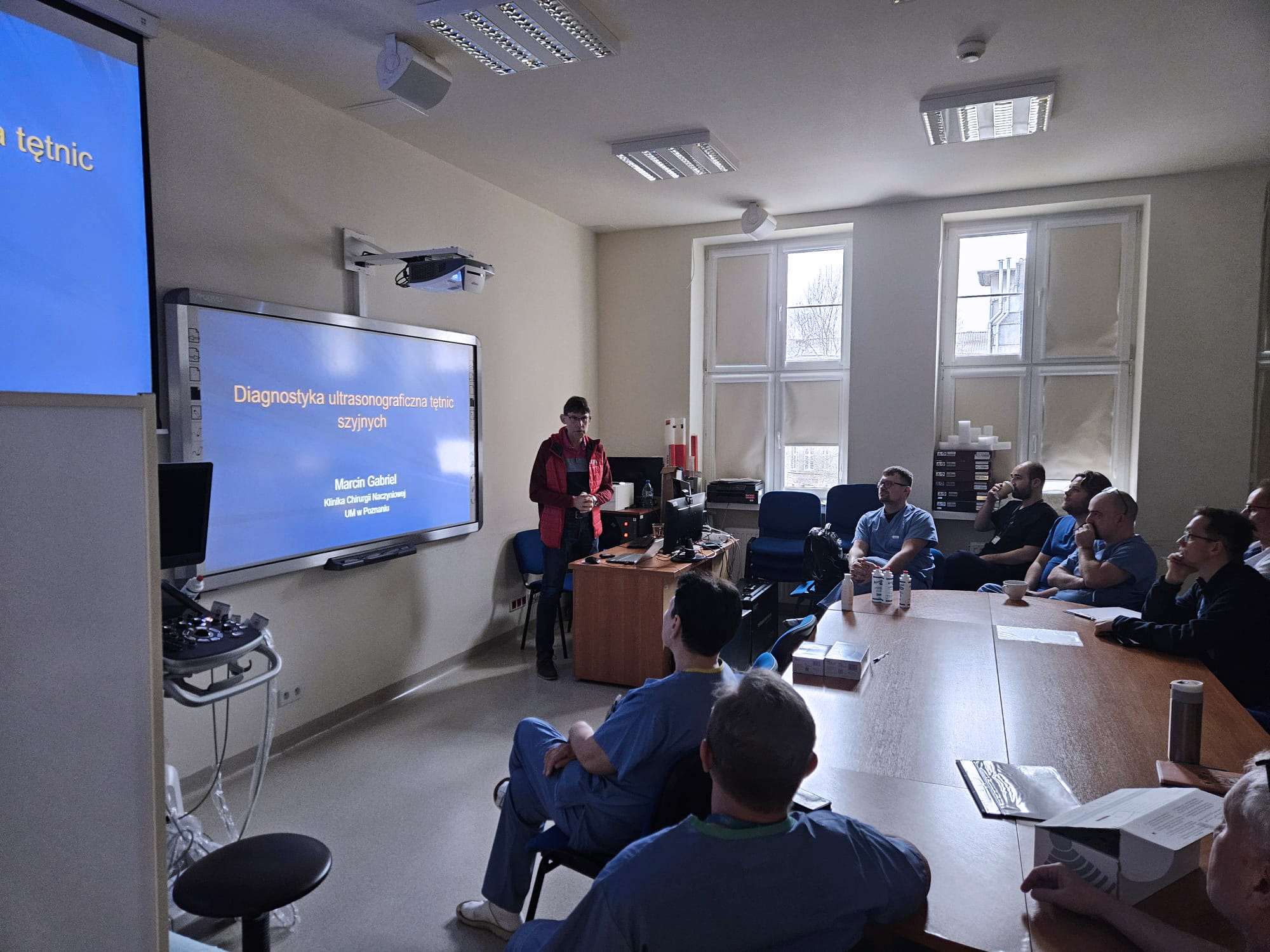
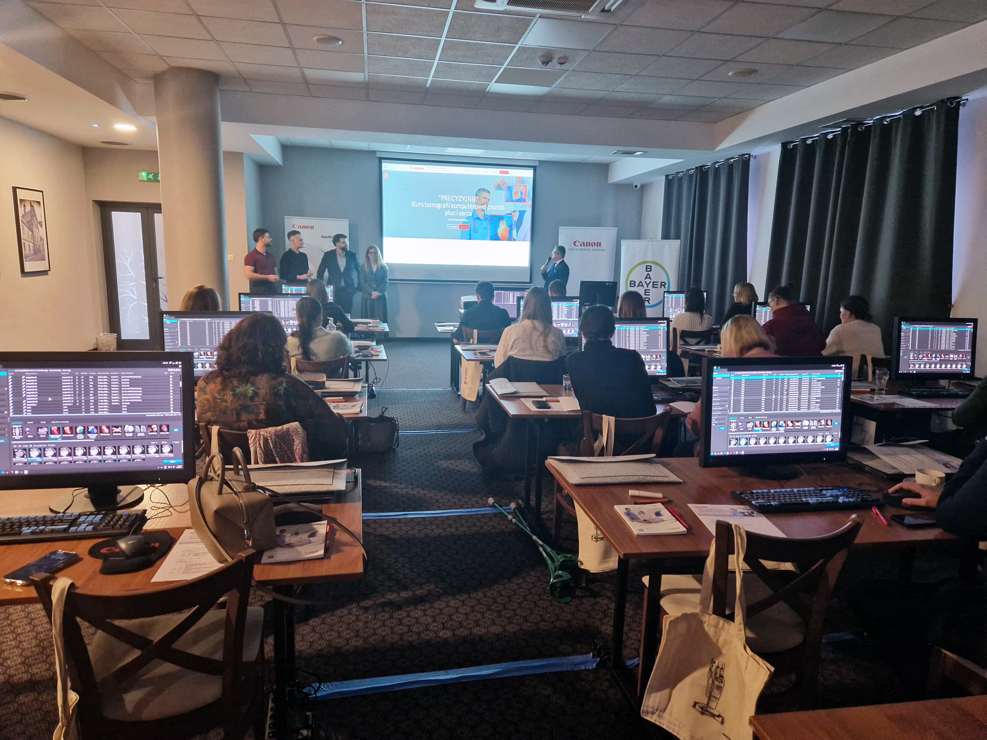
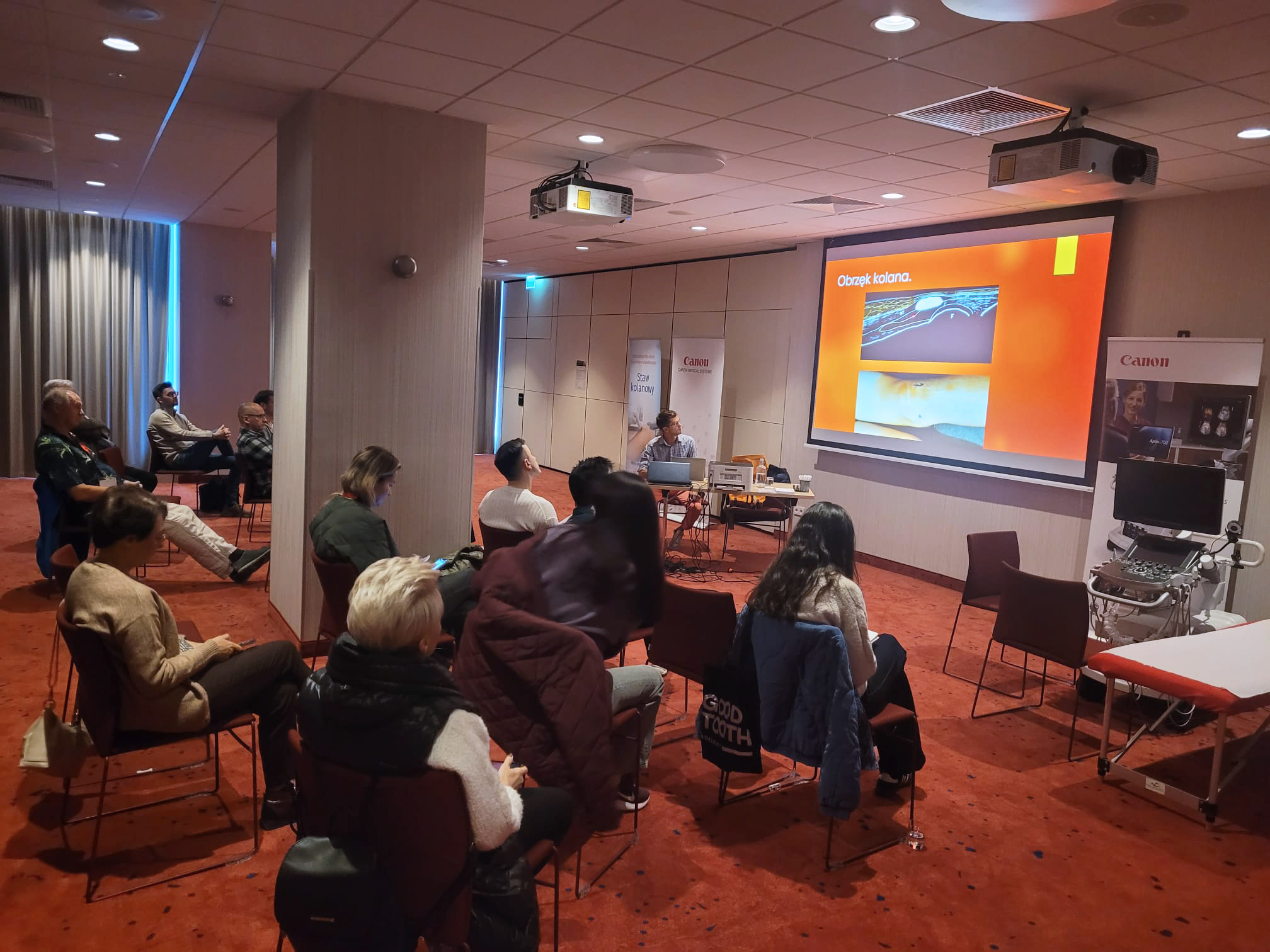
Prowadzący: prof. dr hab. n. med. Marcin Gabriel oraz dr n. med. Jolanta Tomczak
Uniwersytecki Szpital Kliniczny nr 2 PUM w Szczecinie.
🔹 23 stycznia 2026 roku / SZCZECIN
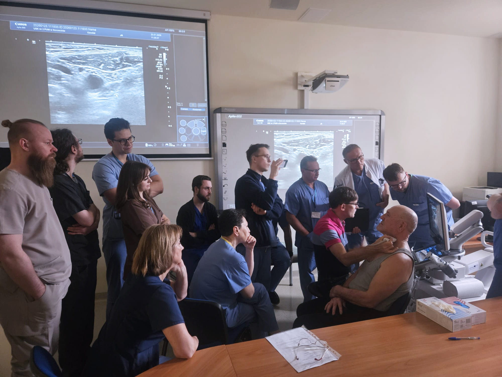
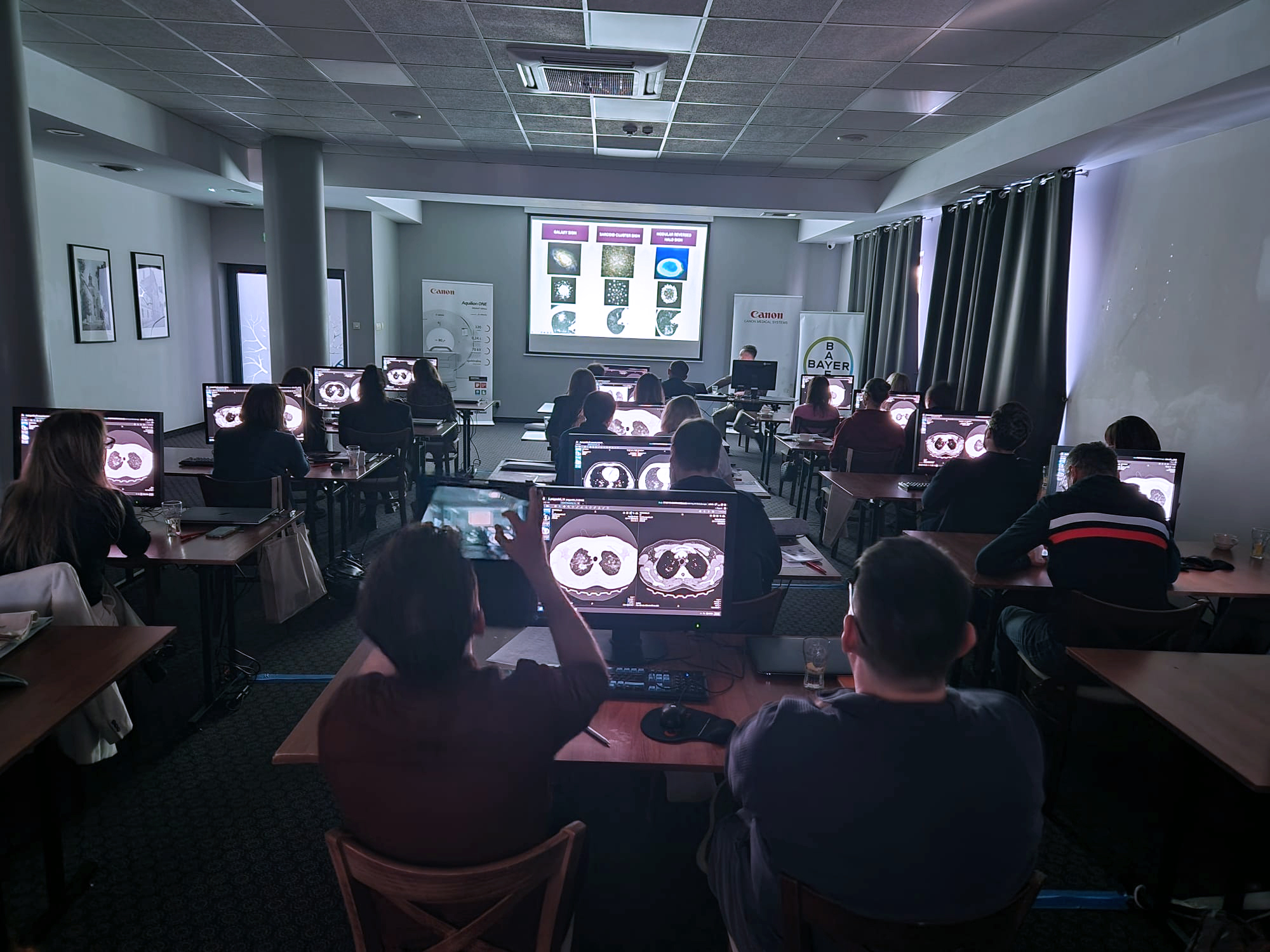
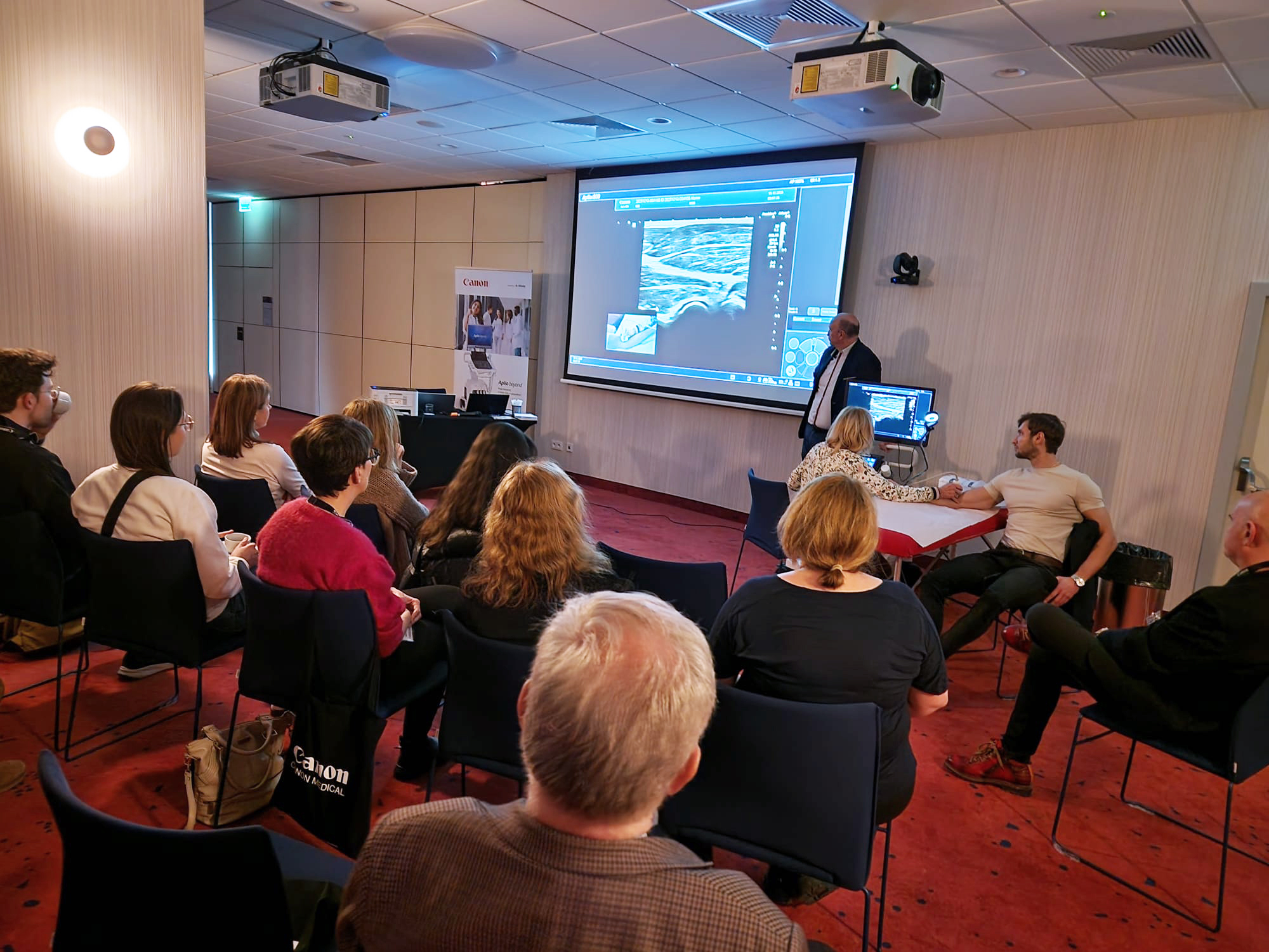

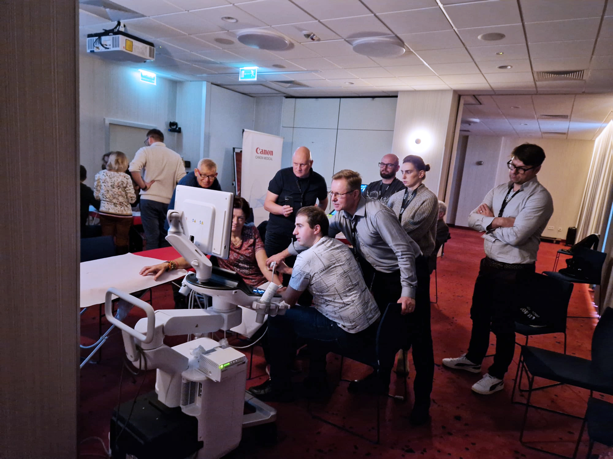
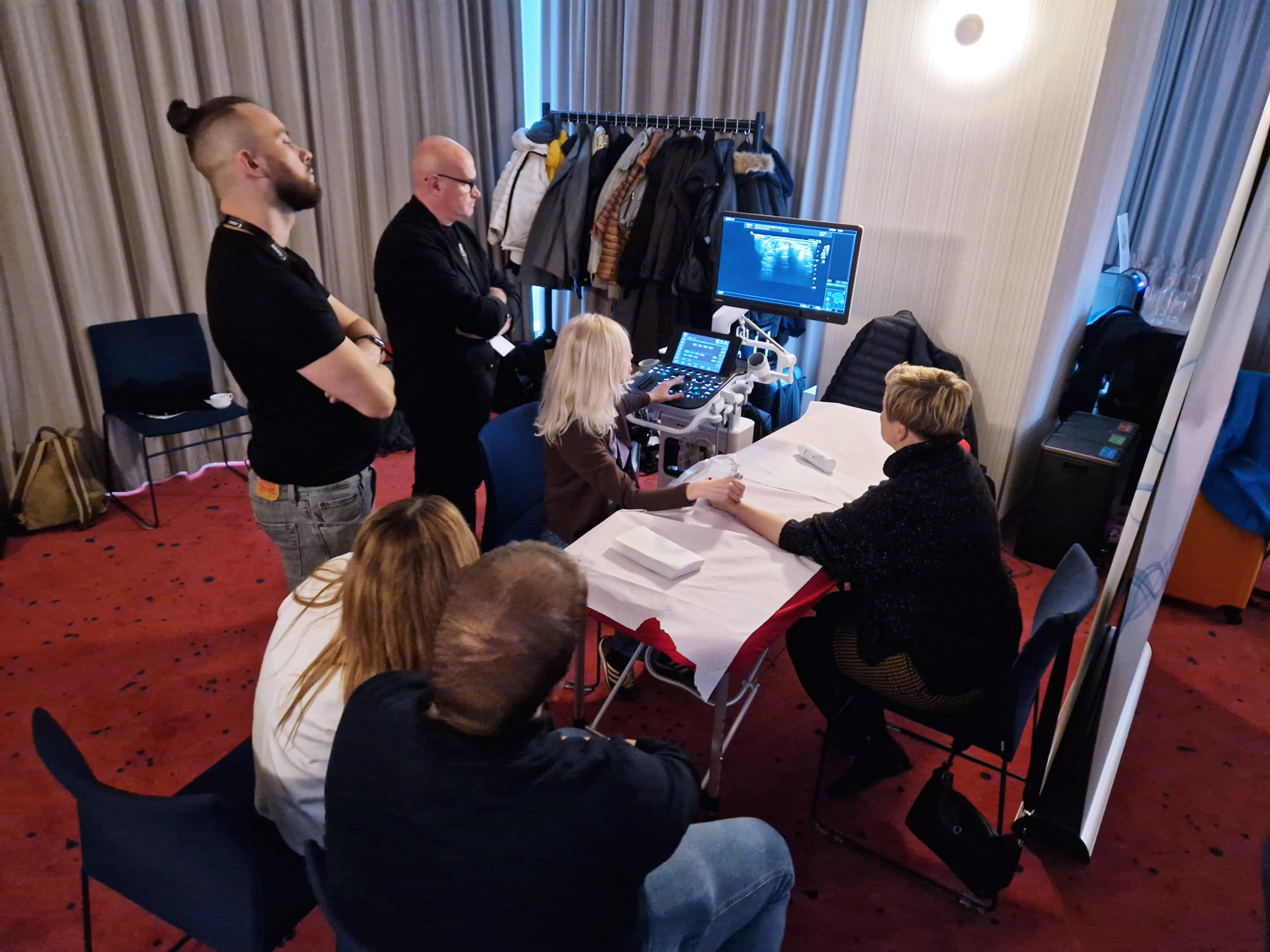
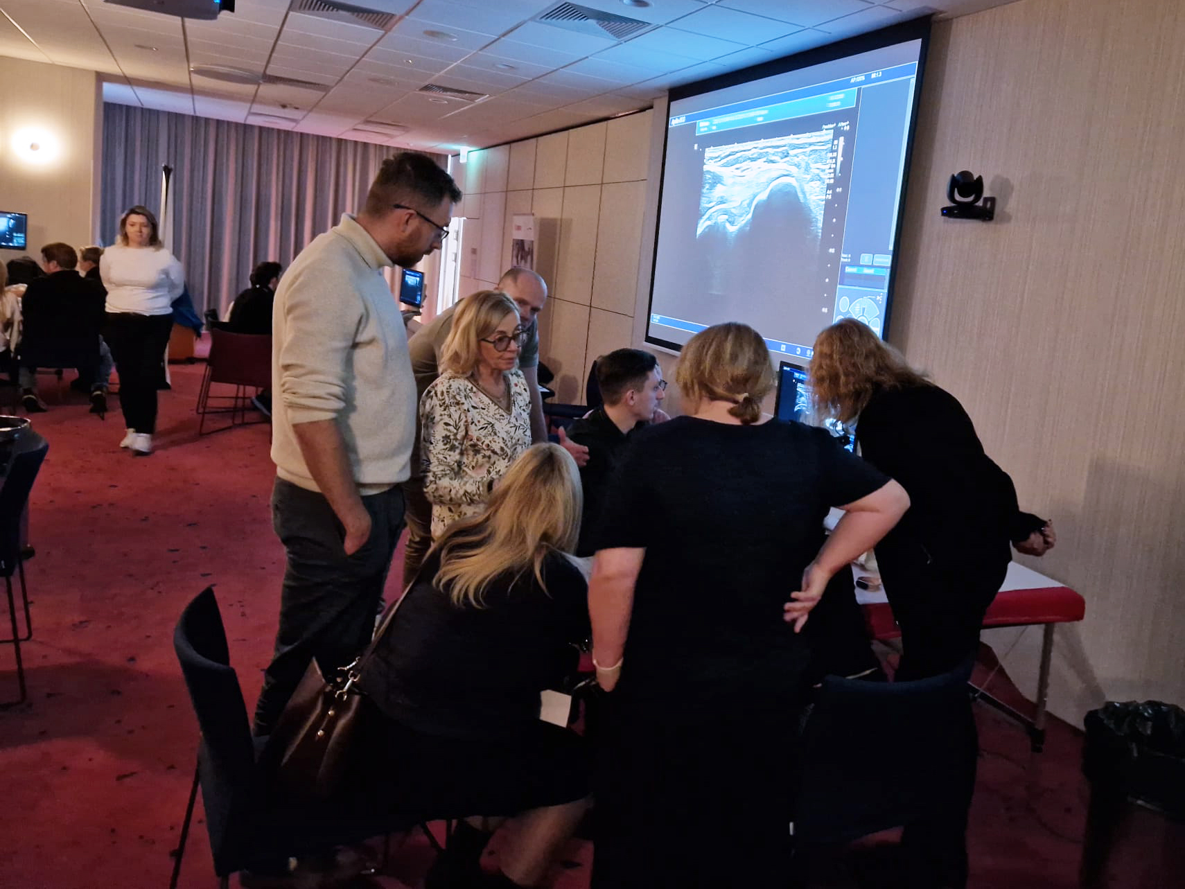
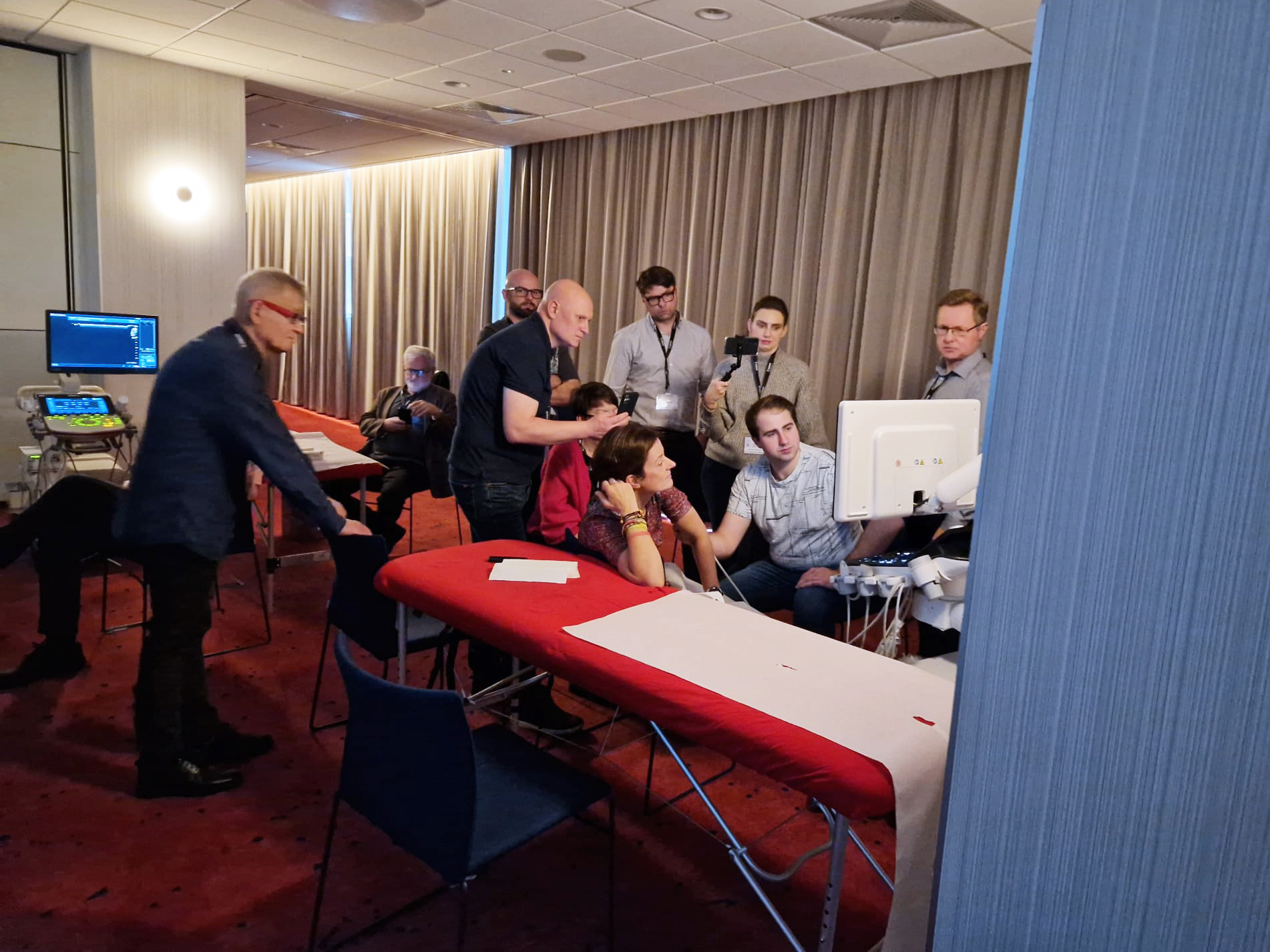
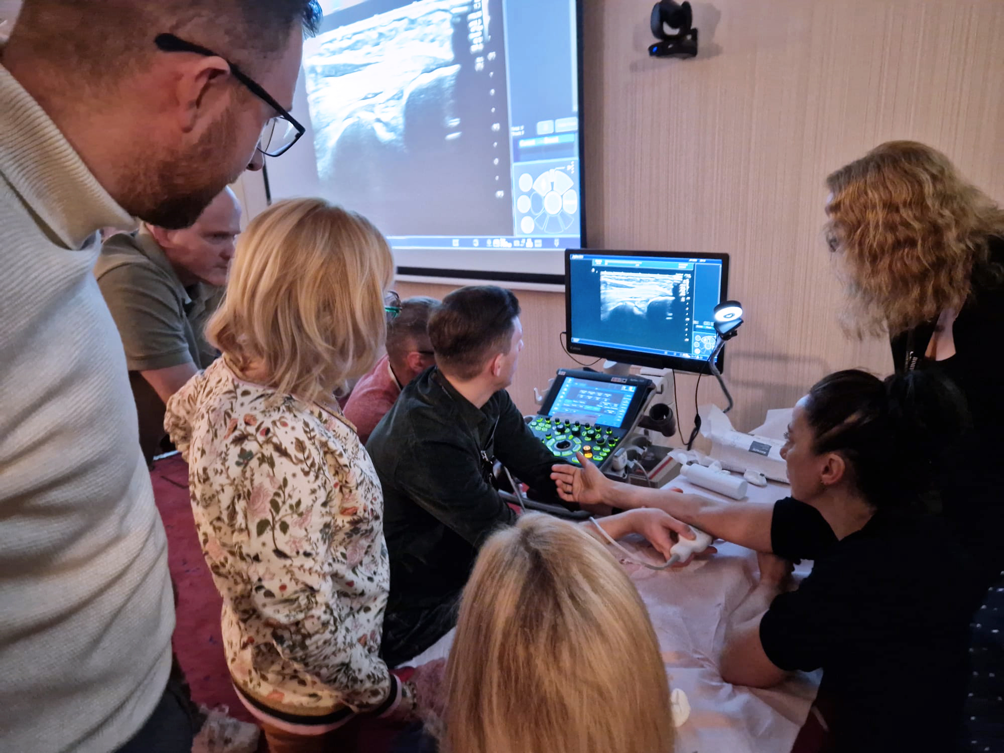
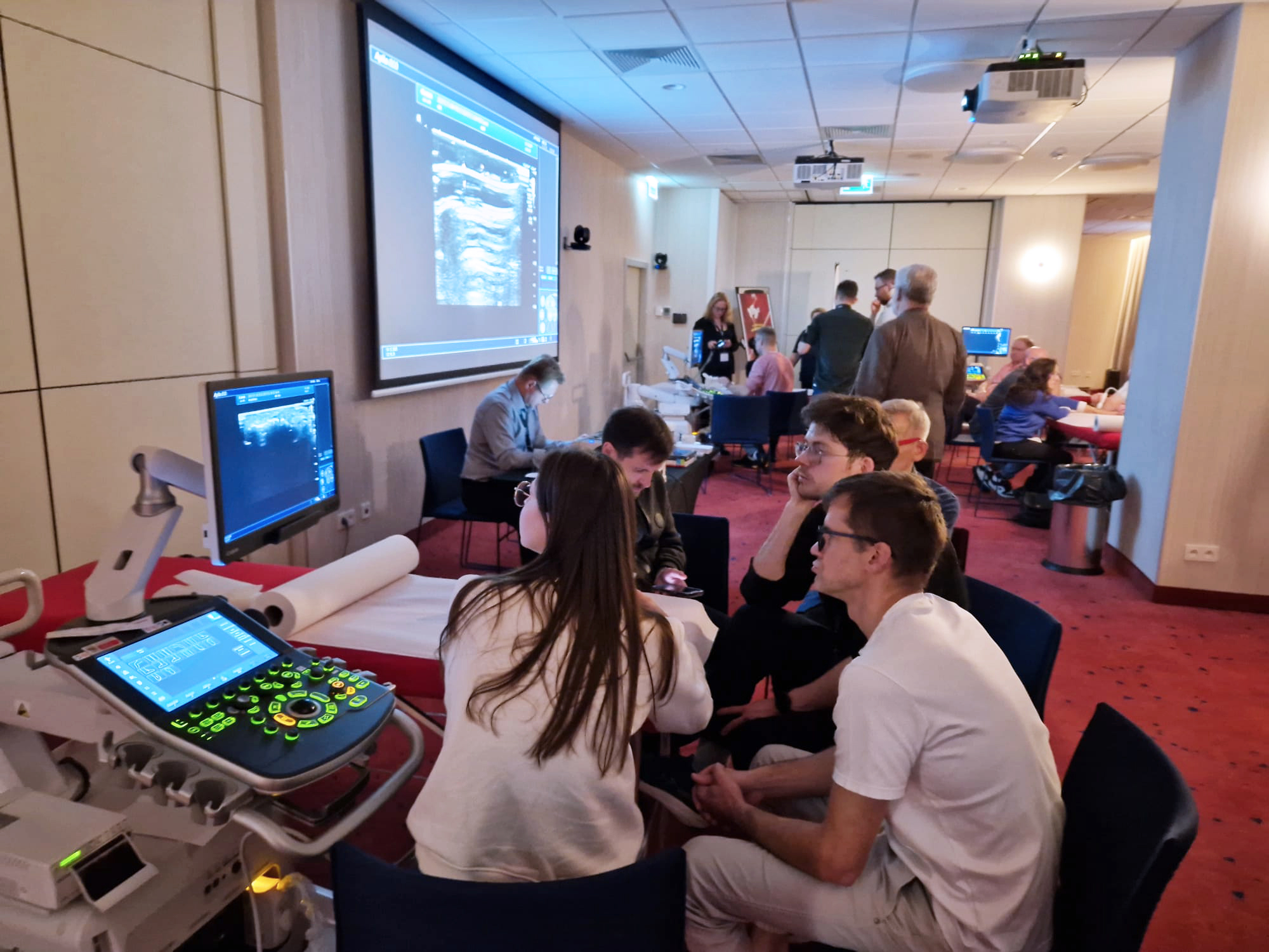
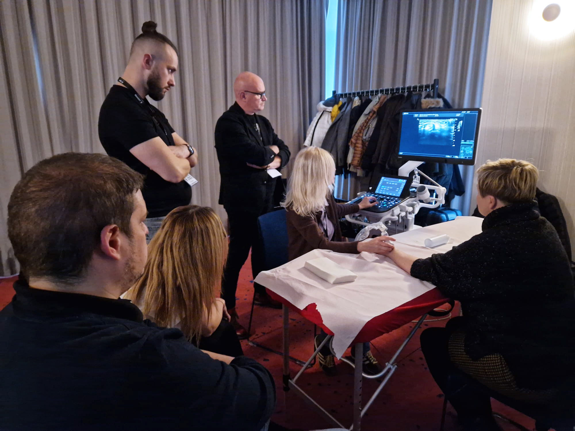


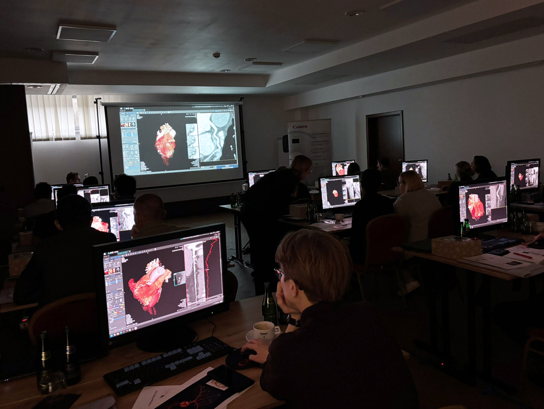
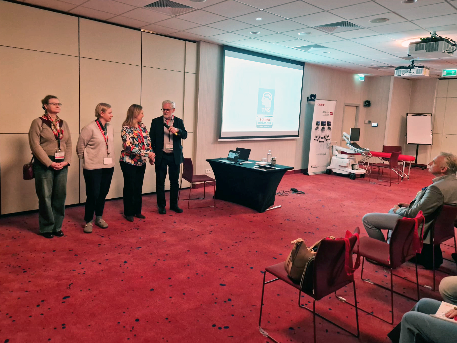
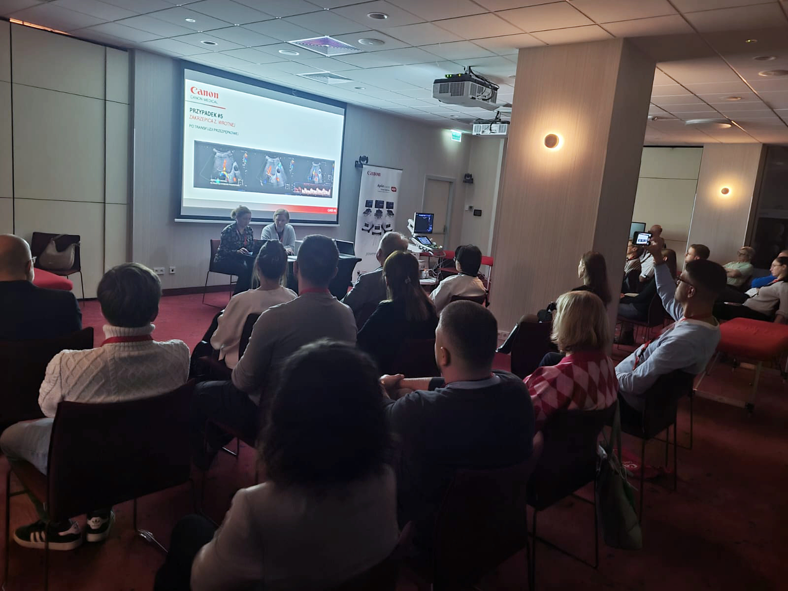
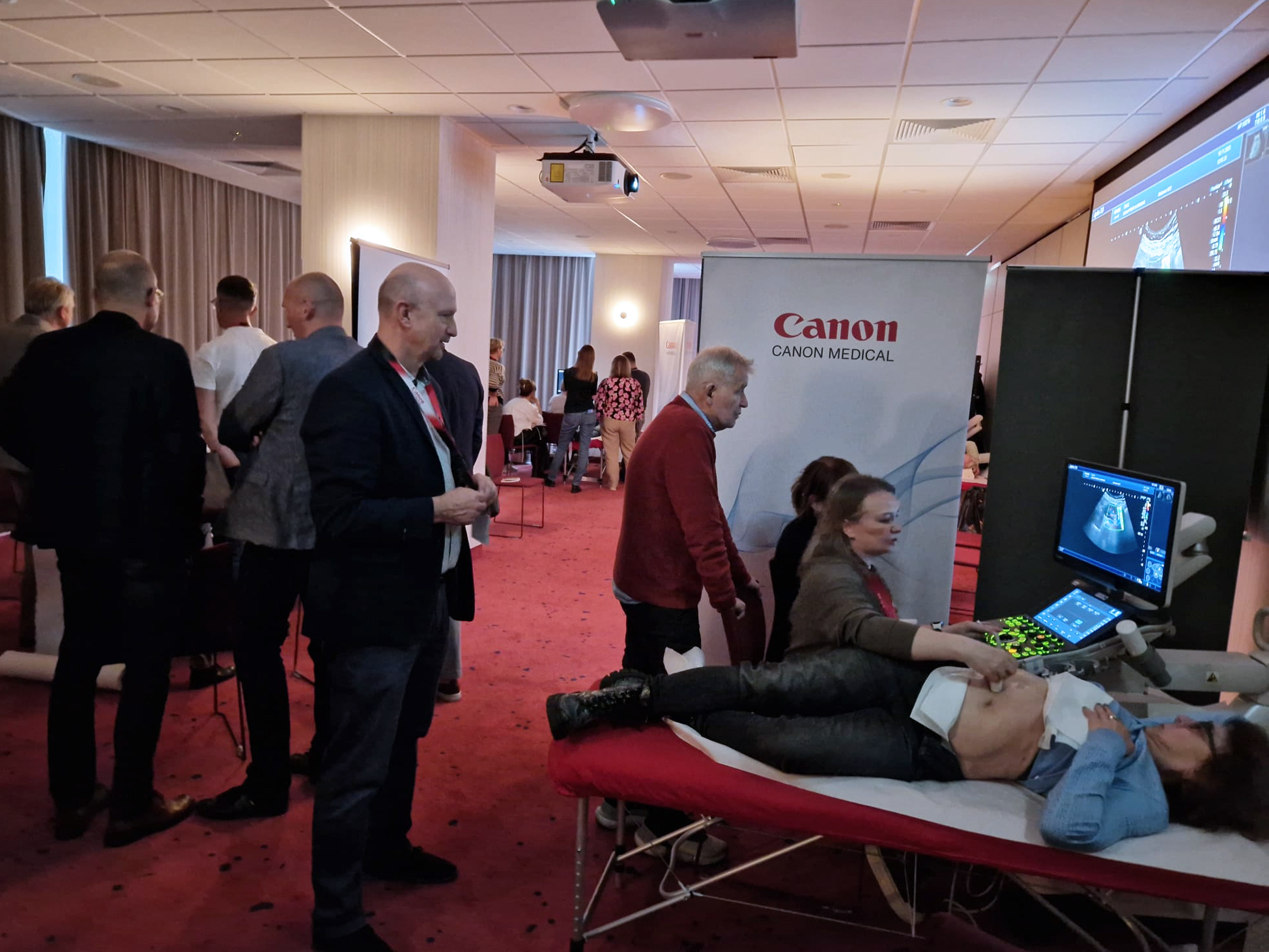
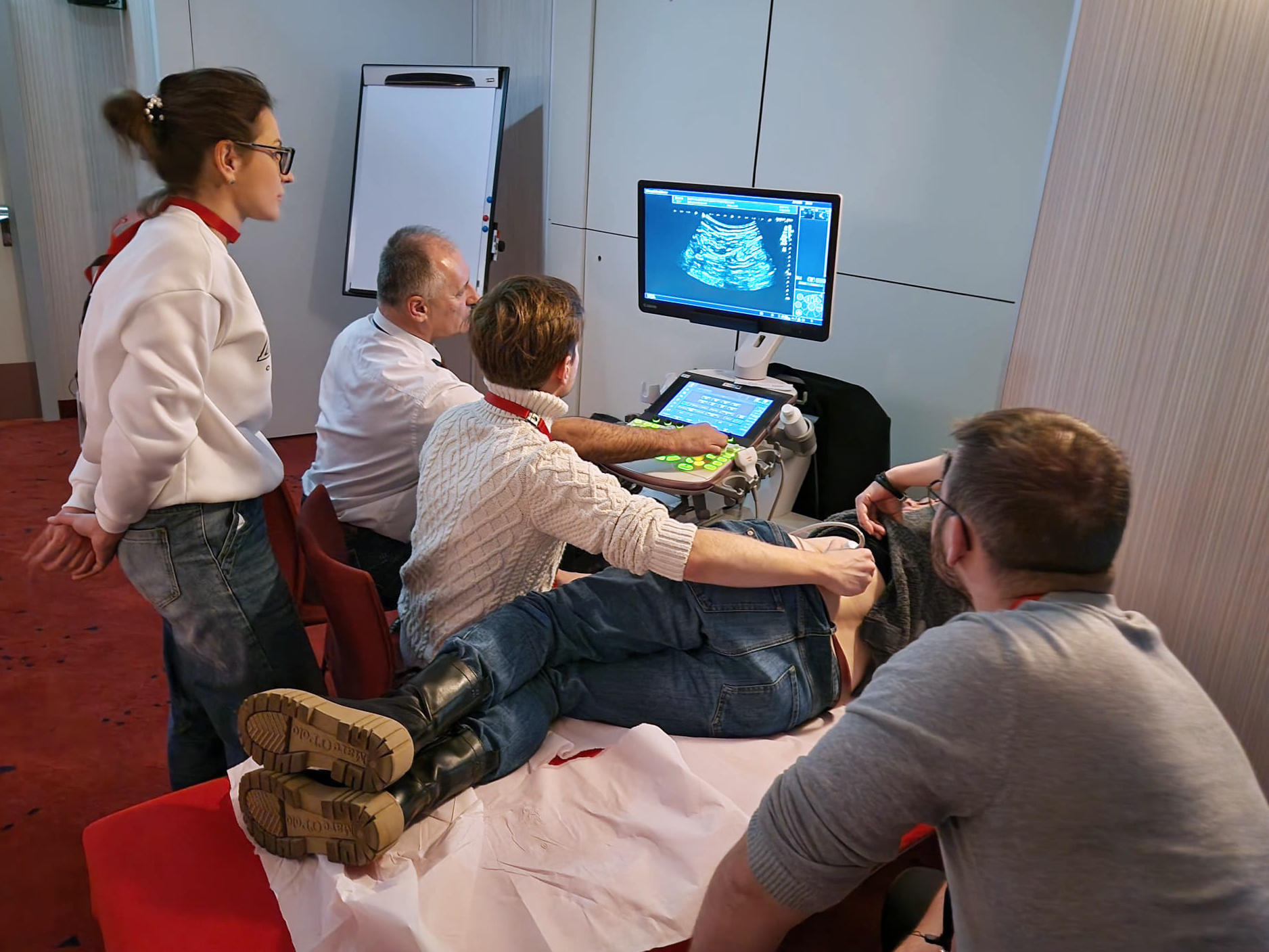
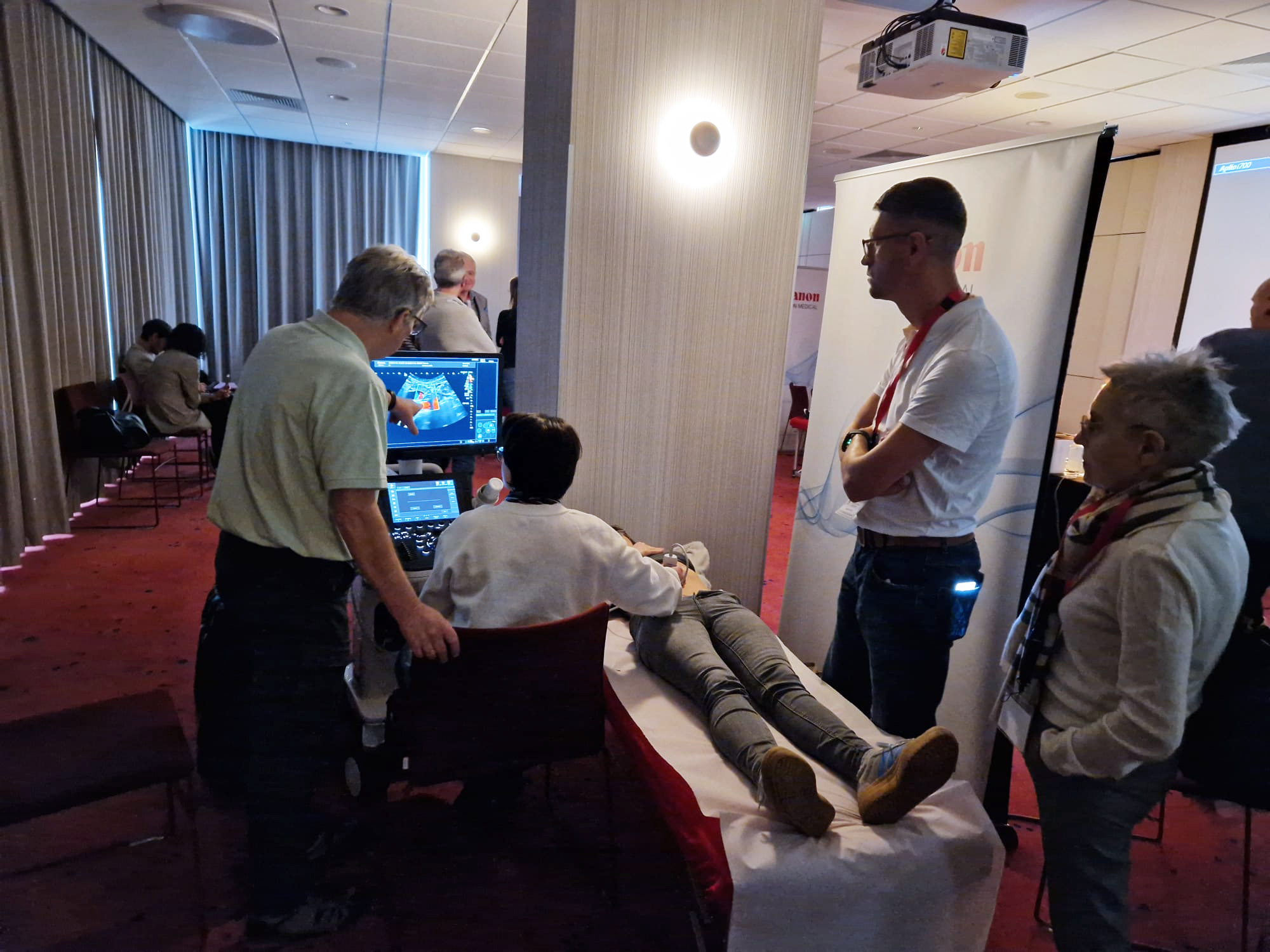
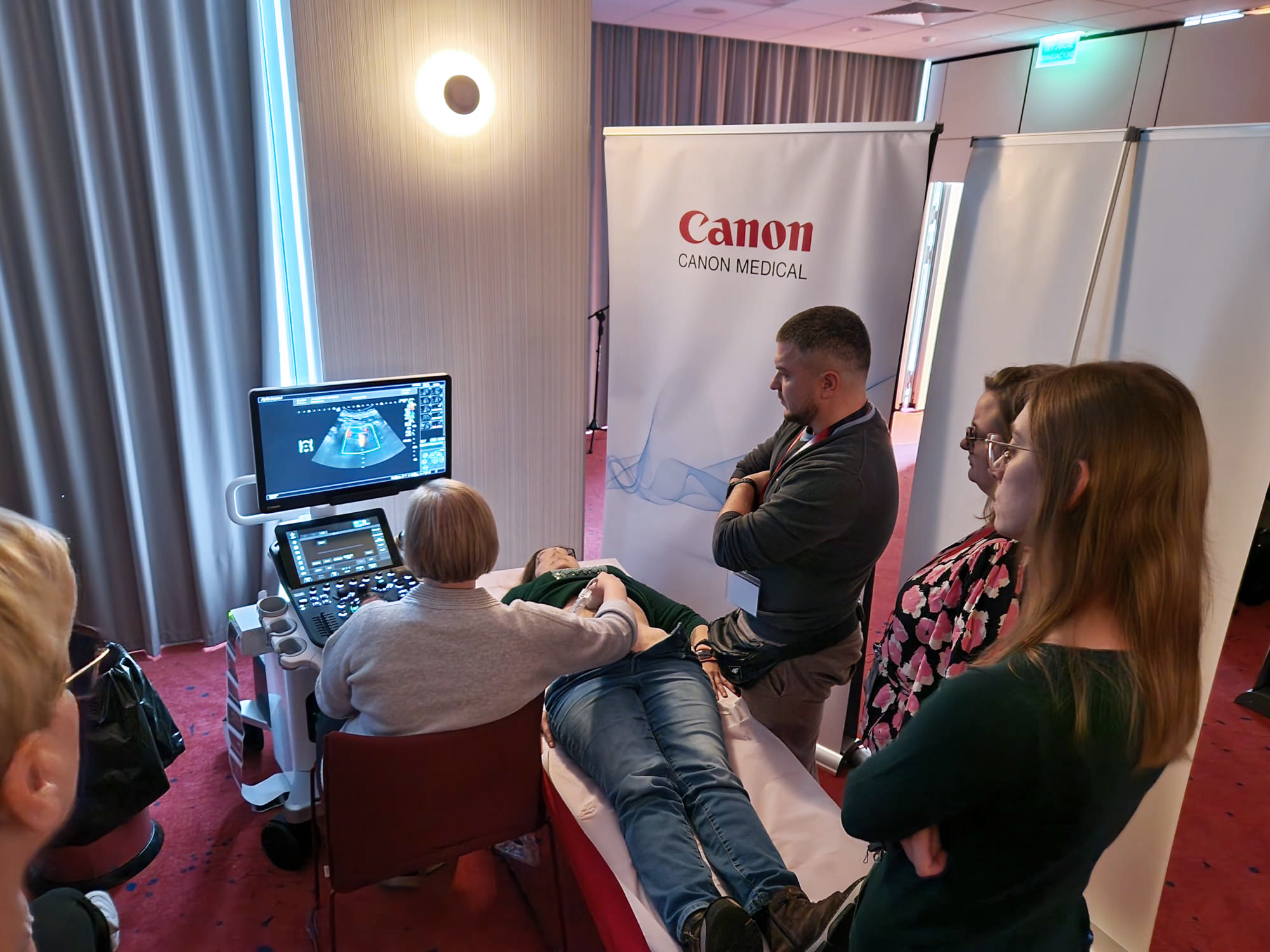
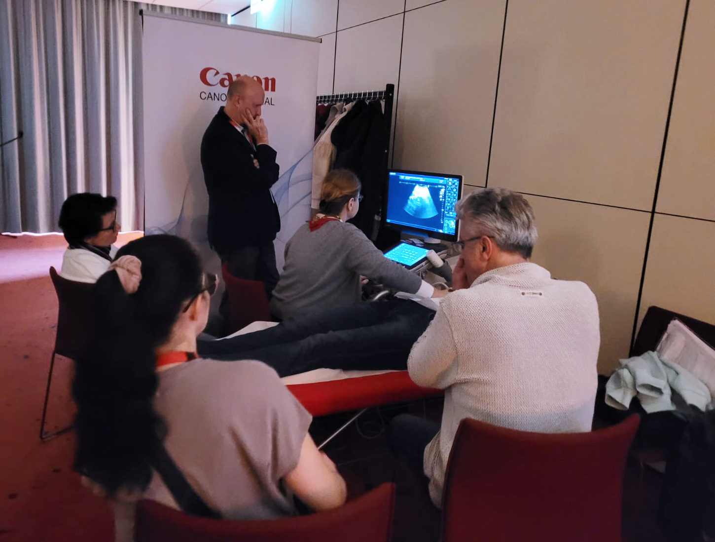
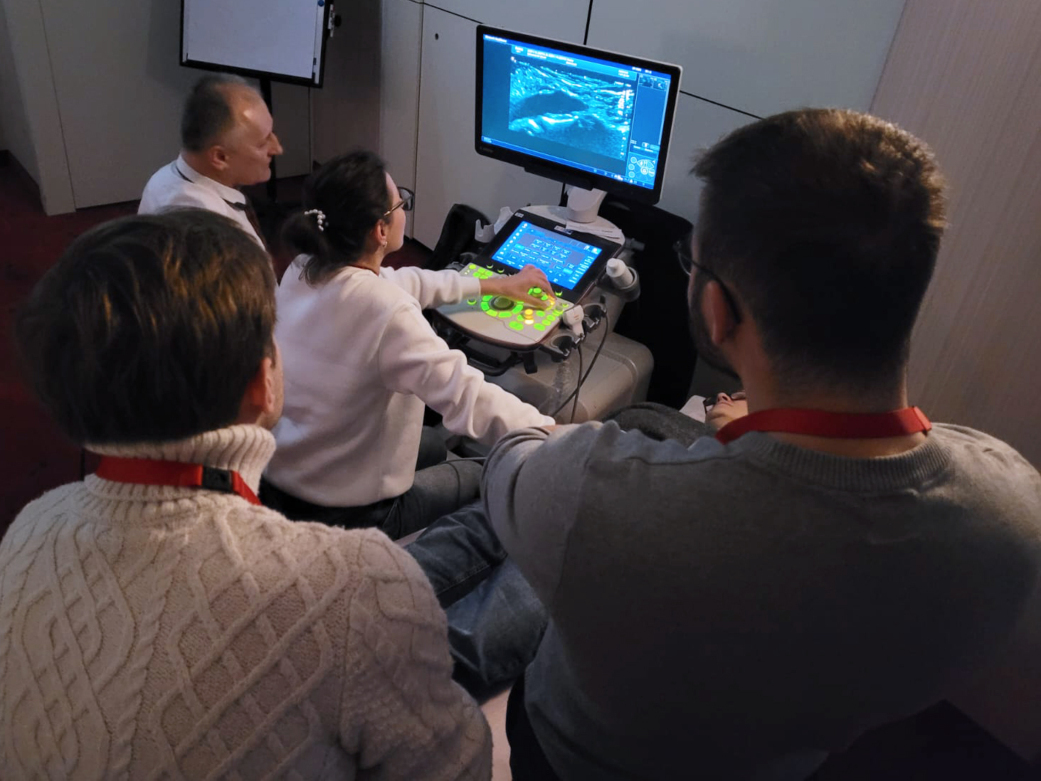
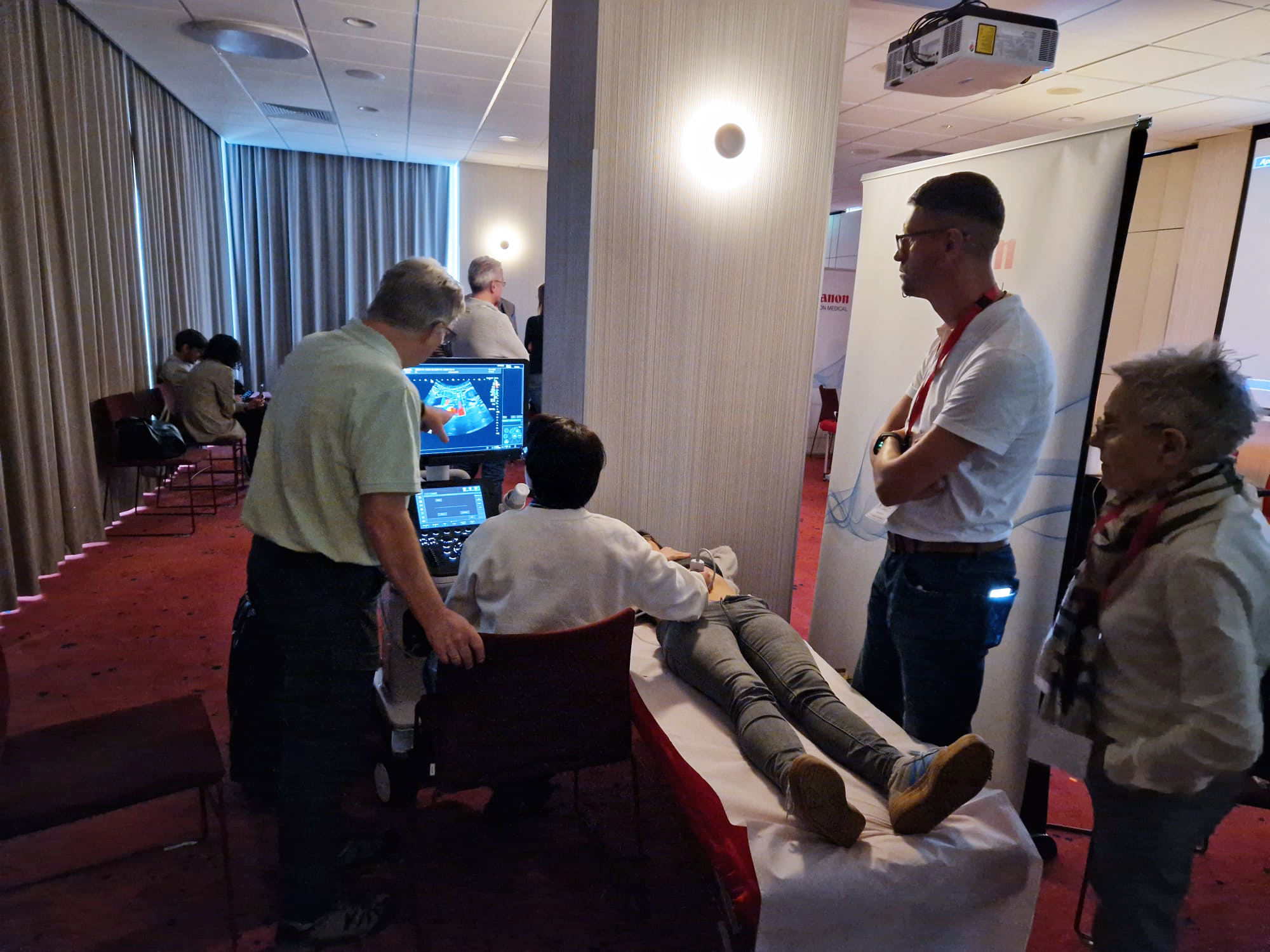
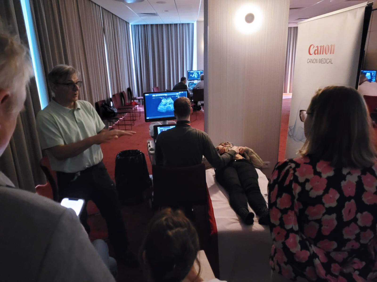


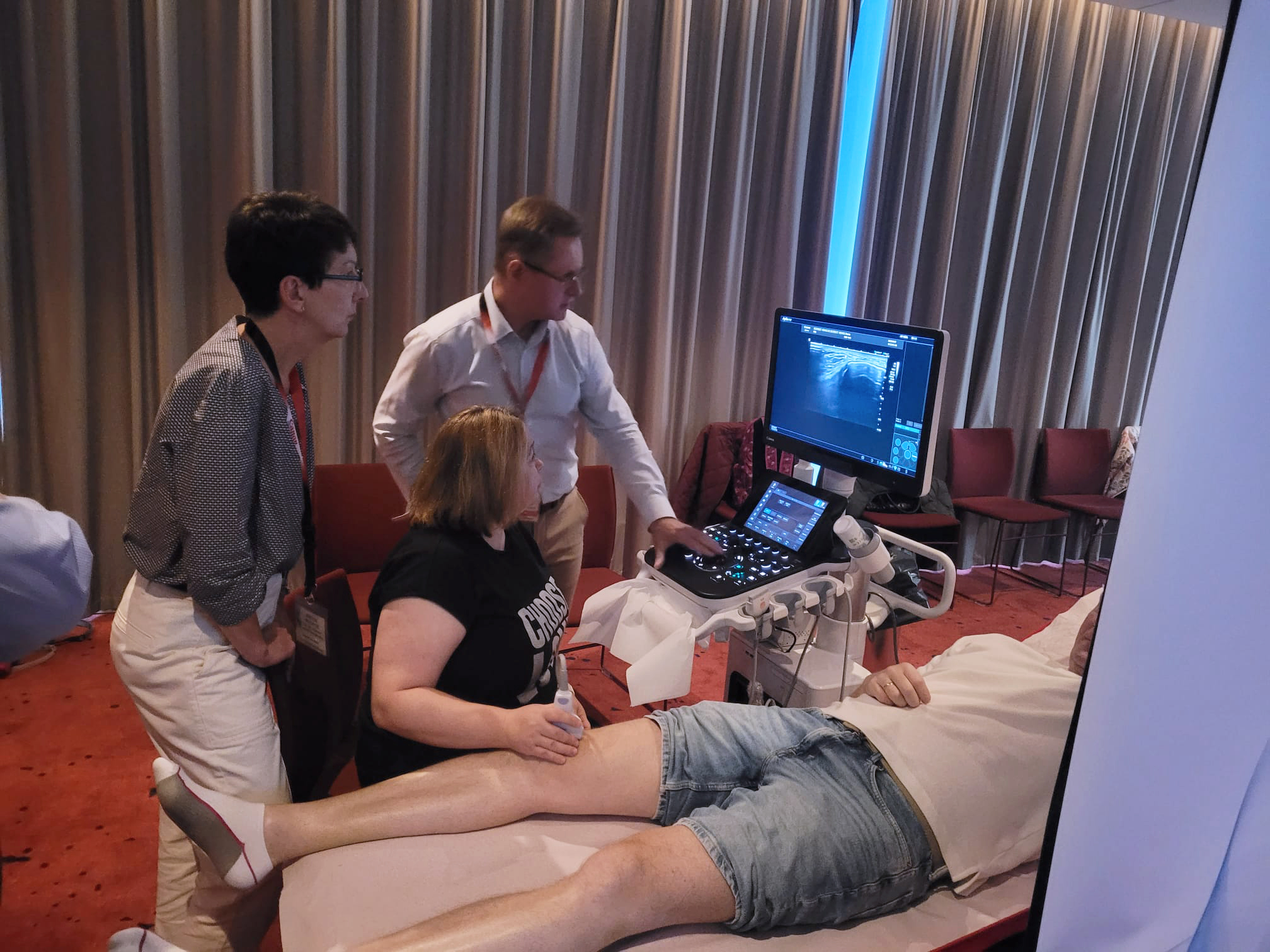
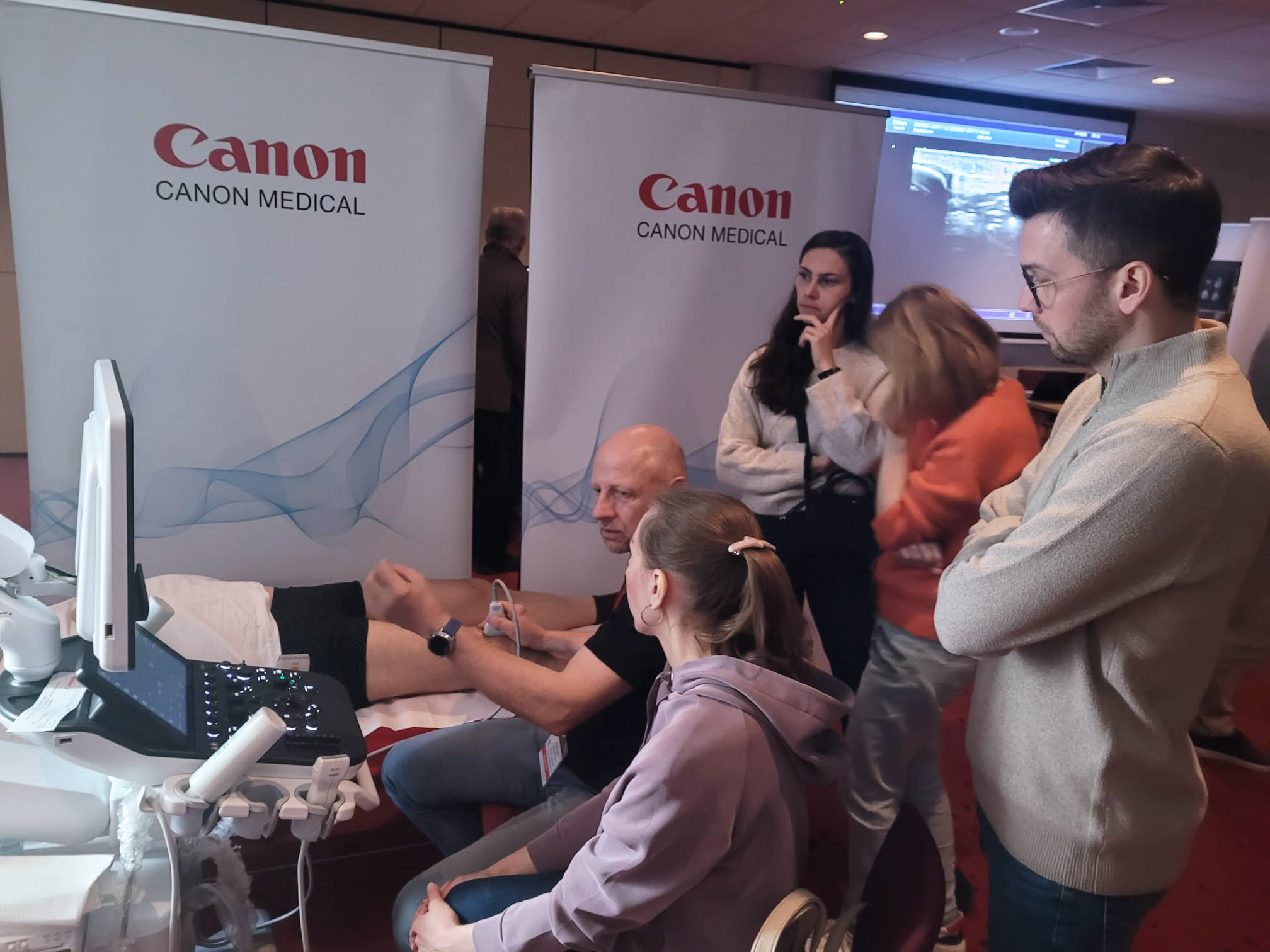
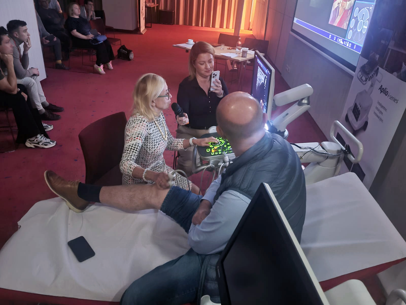
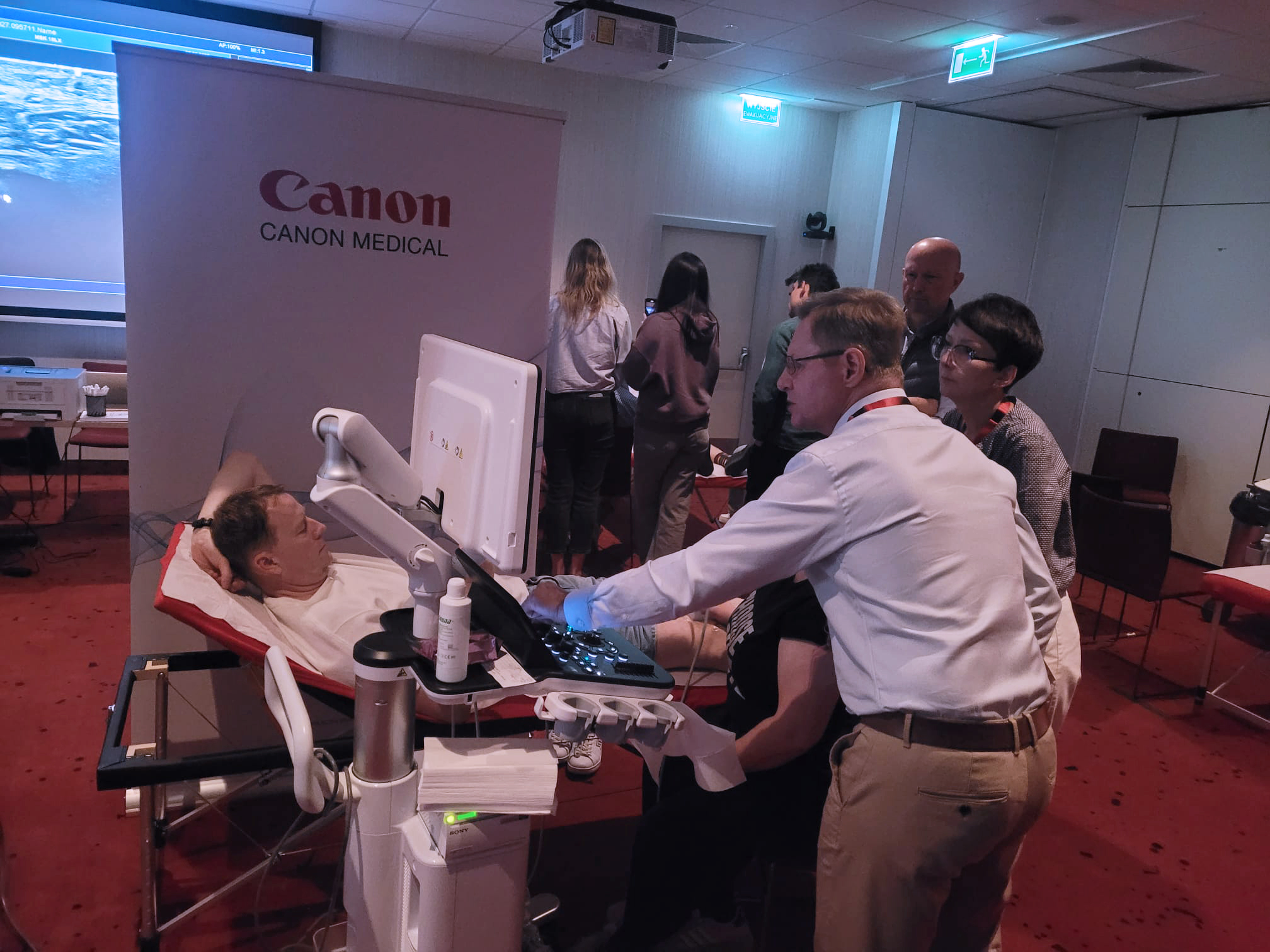
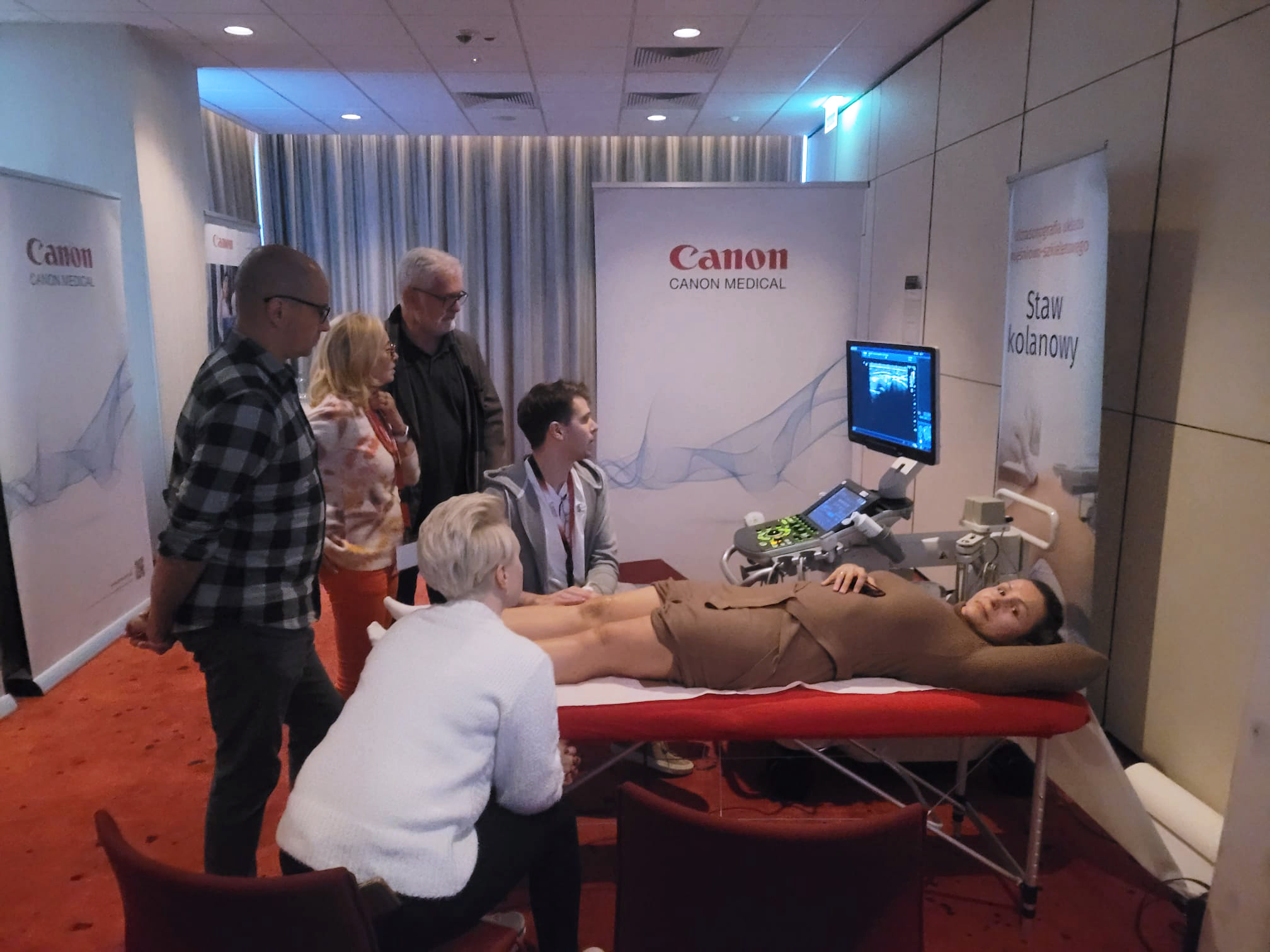
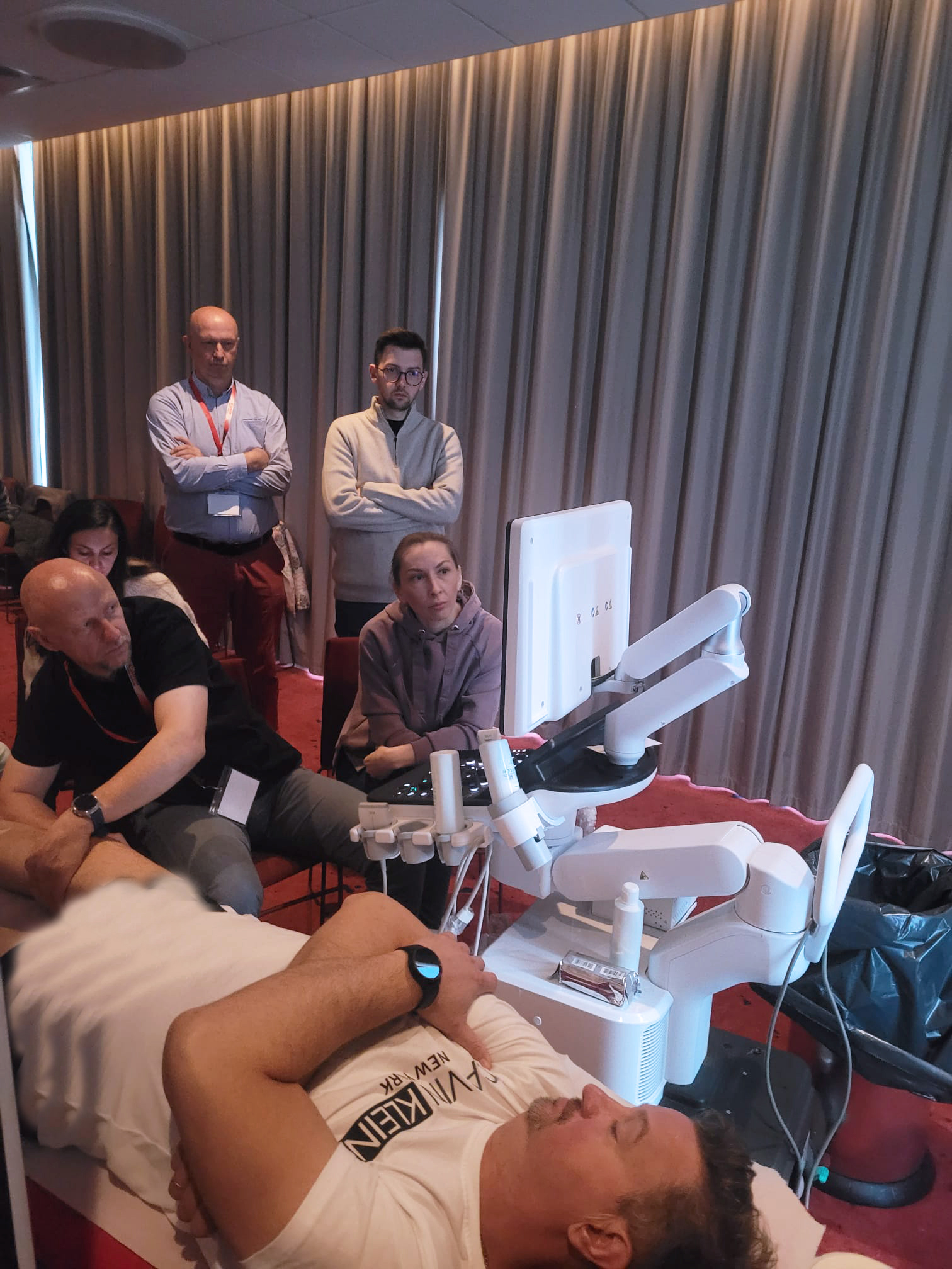
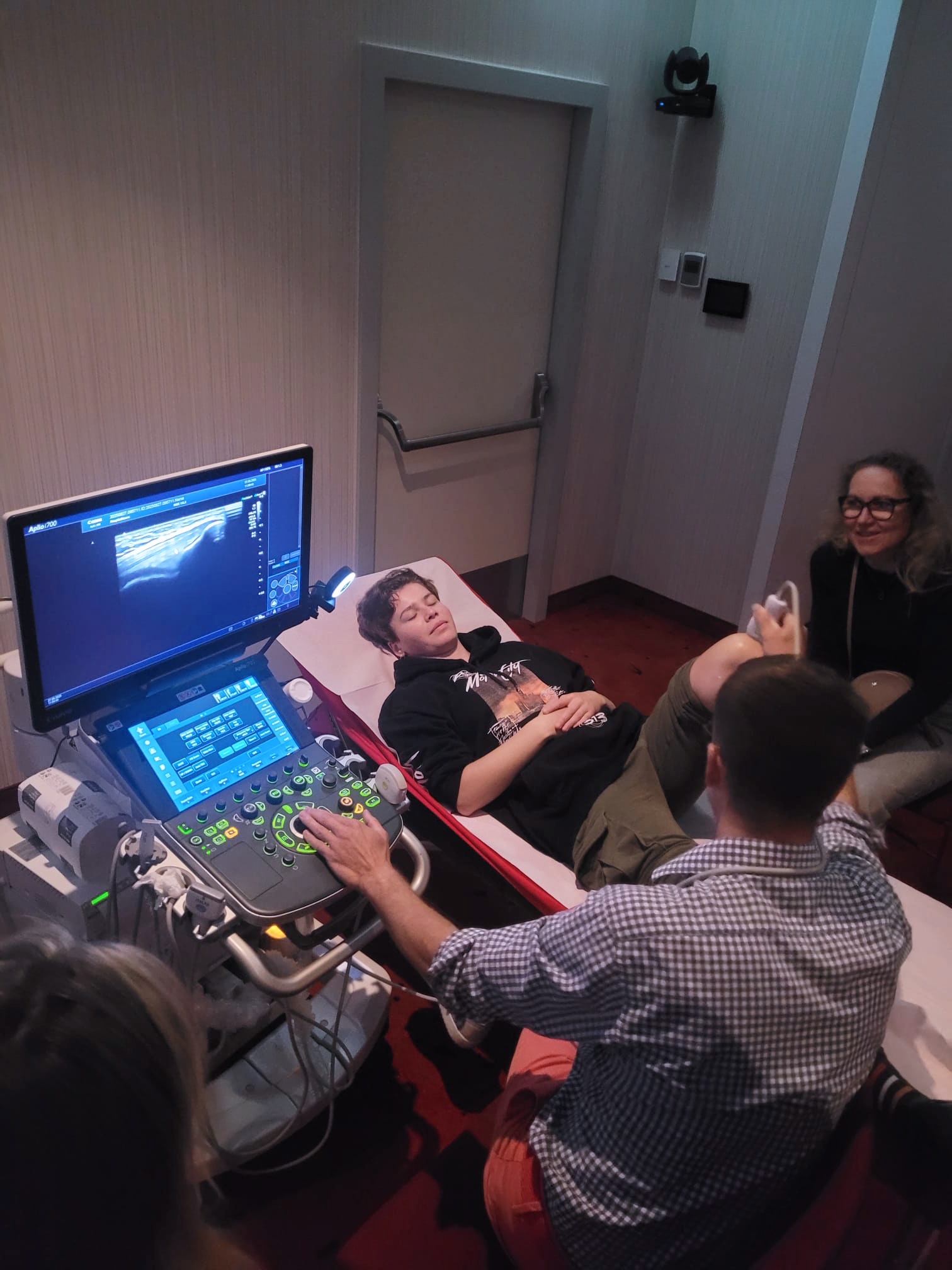
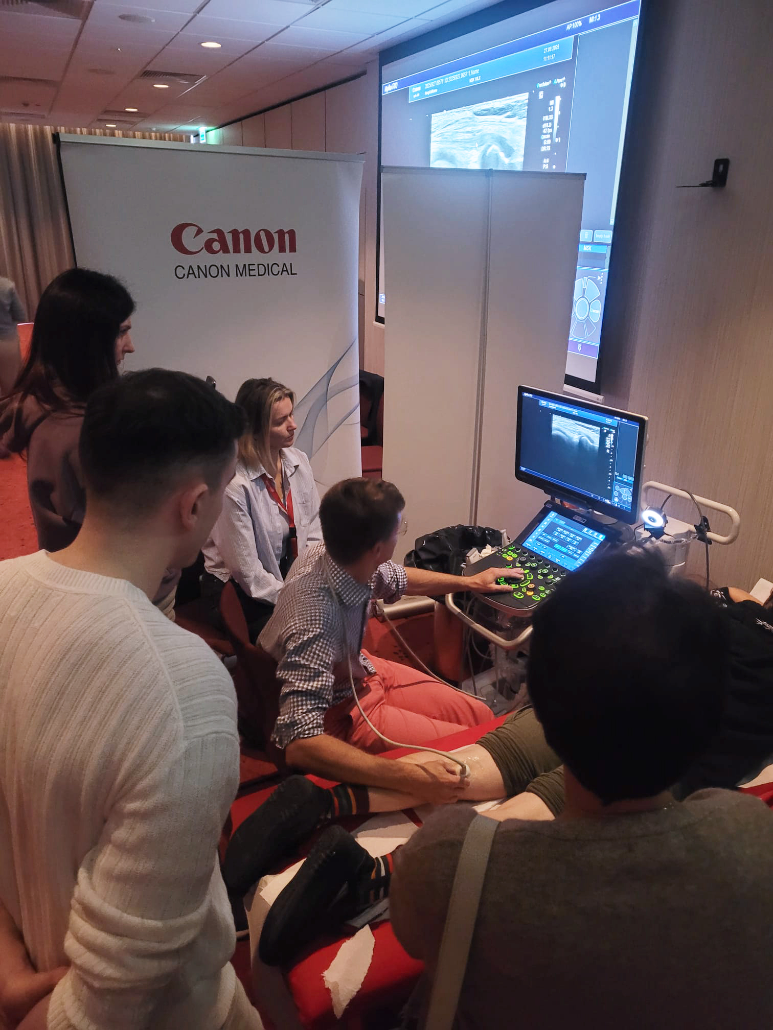
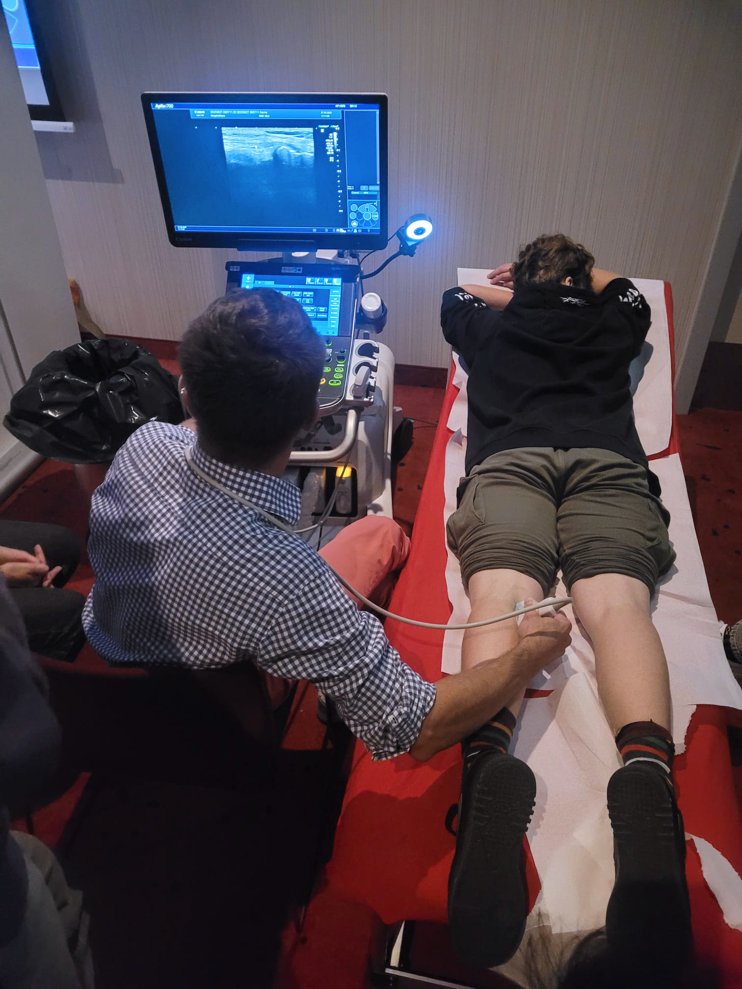
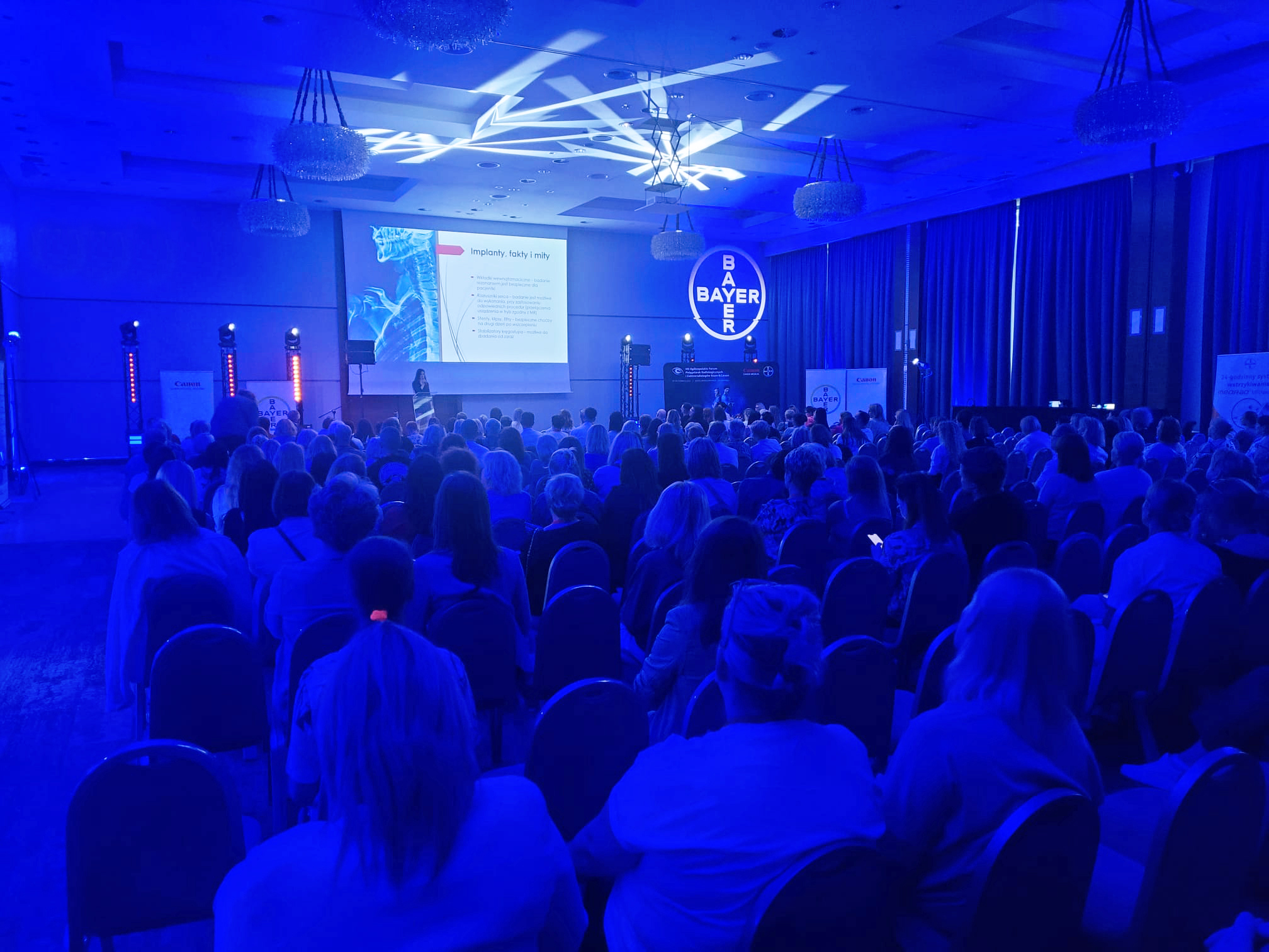
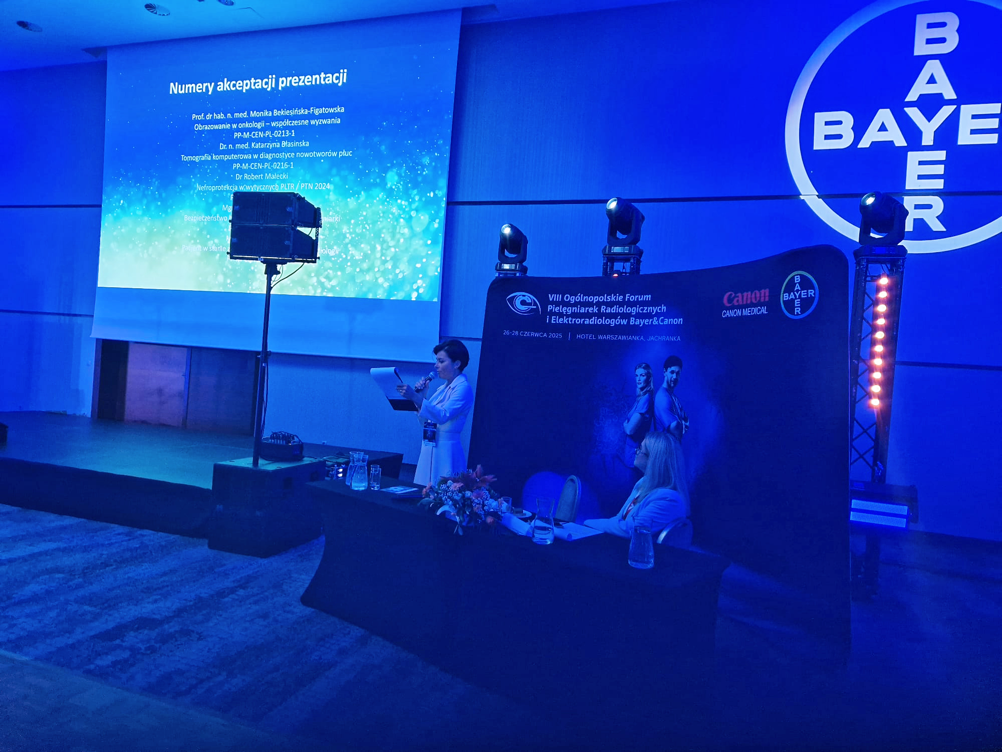
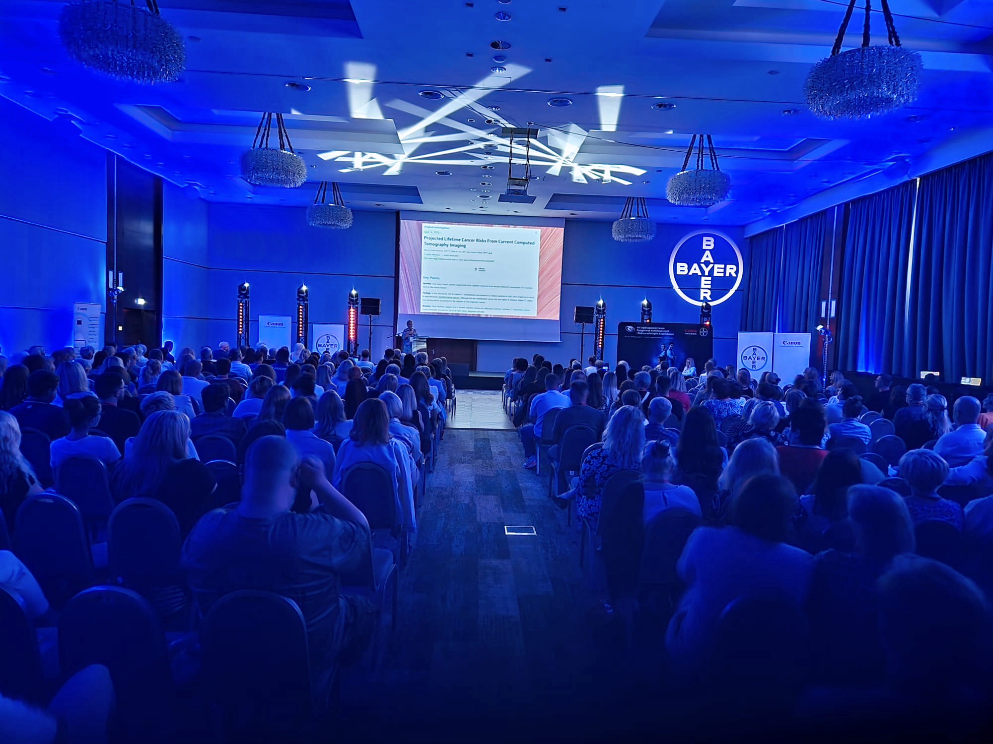
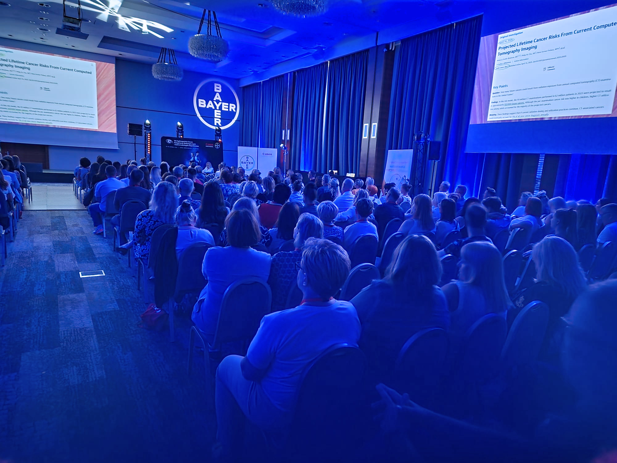
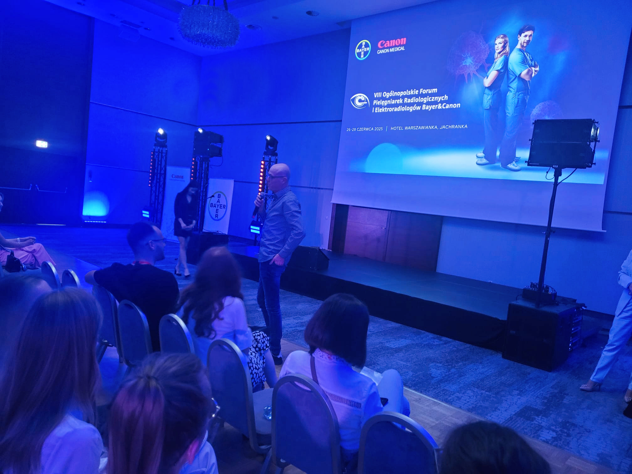
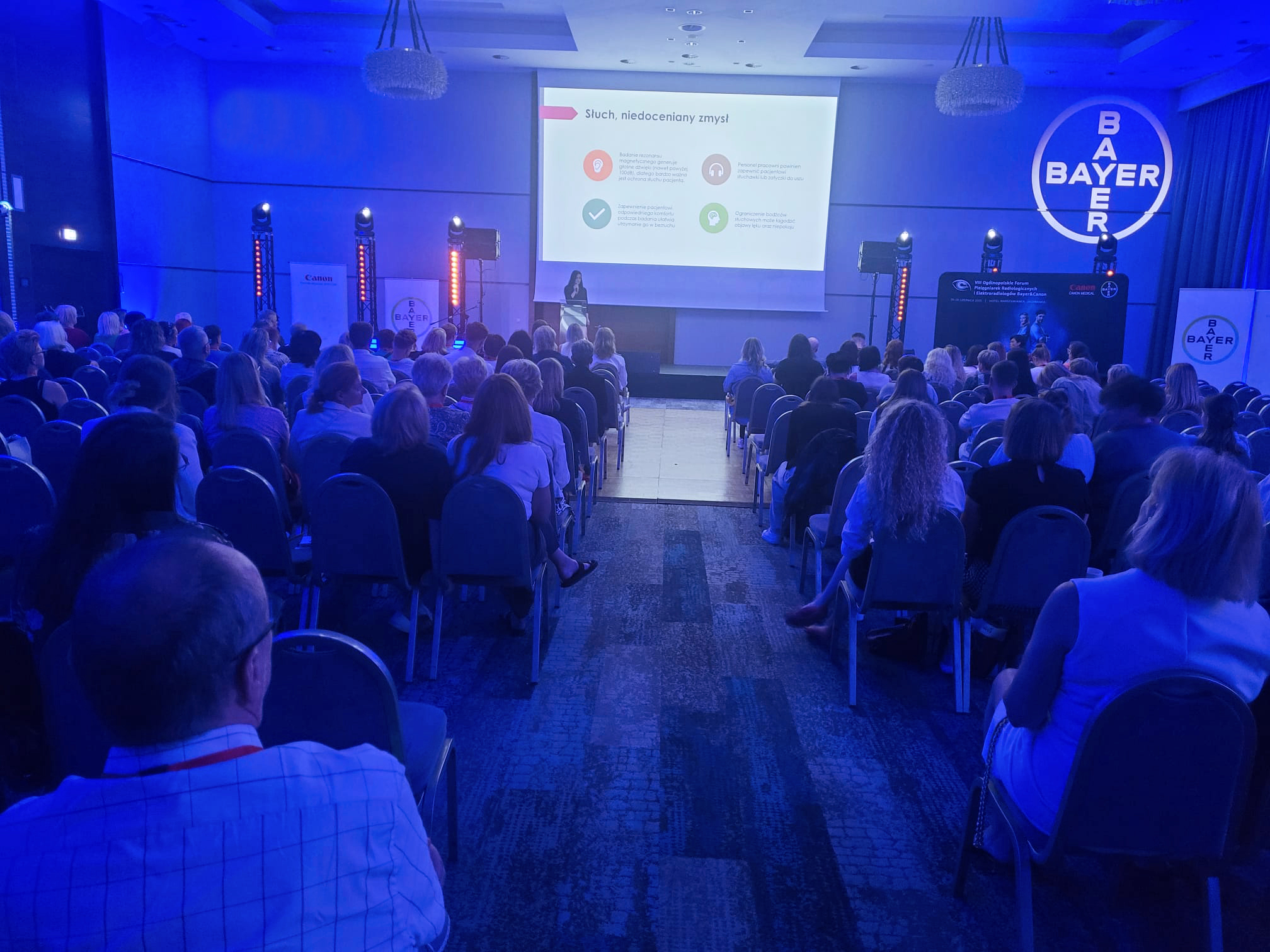
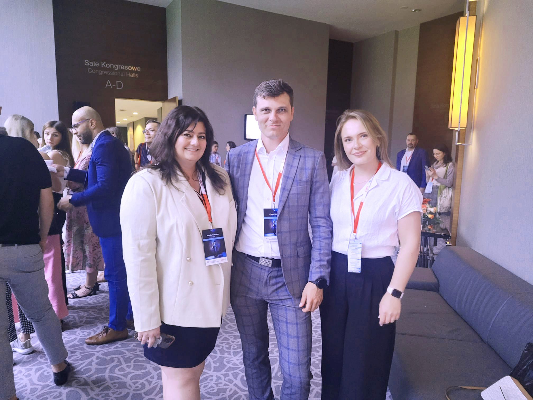
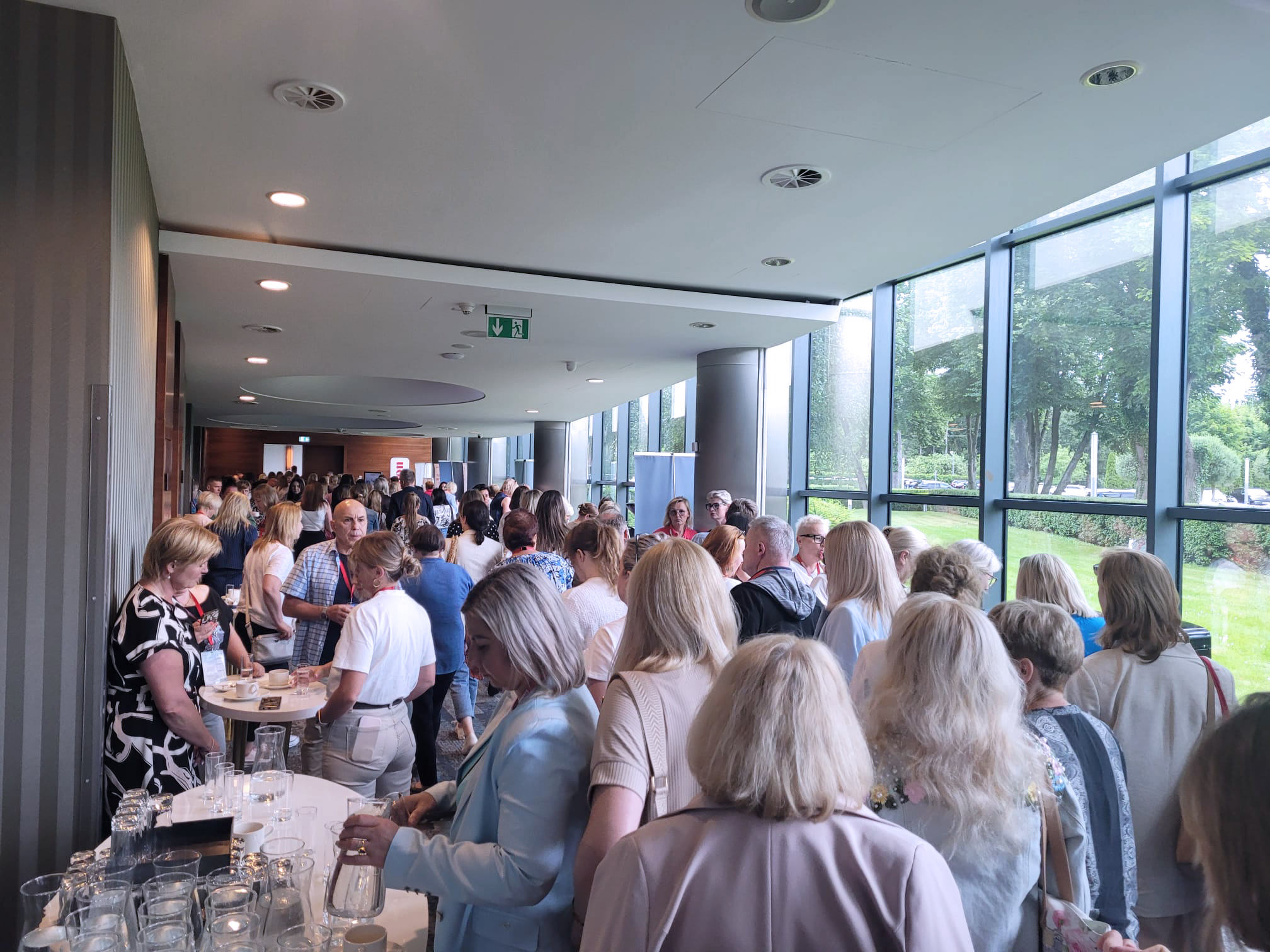
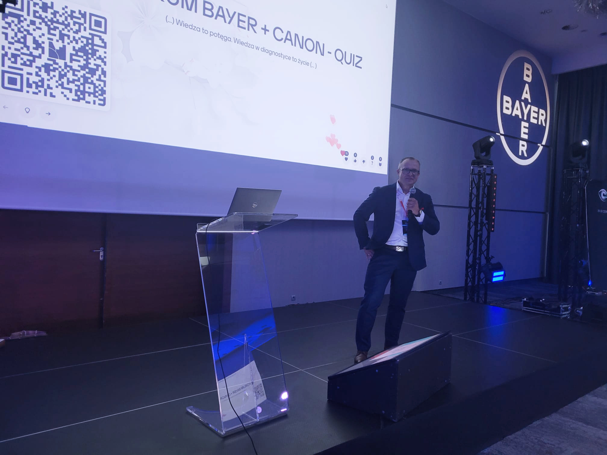
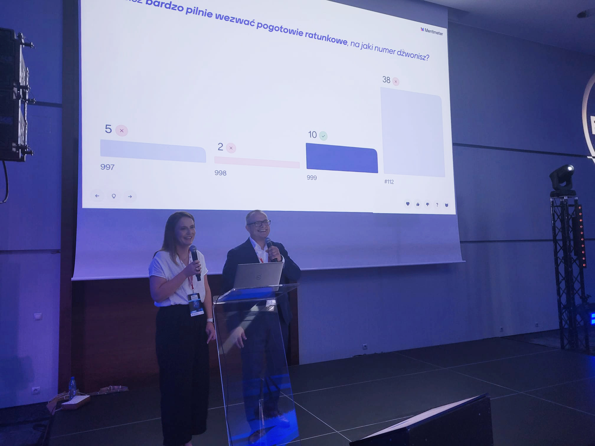
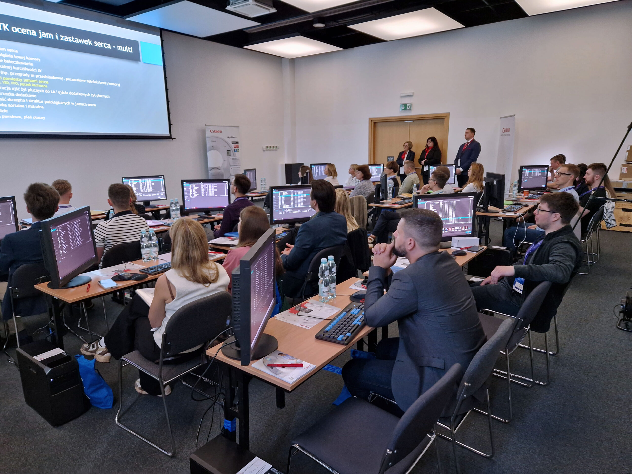
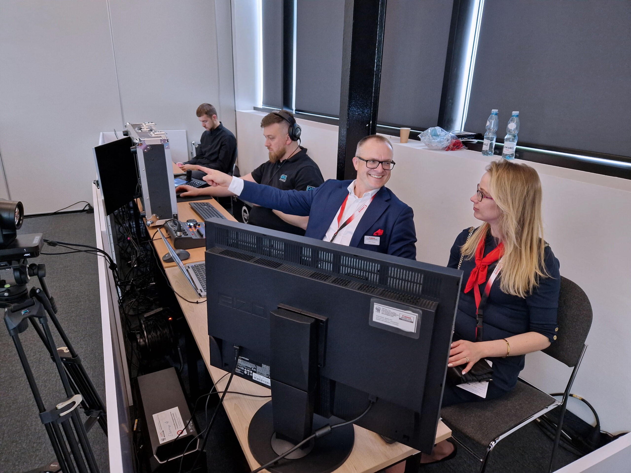
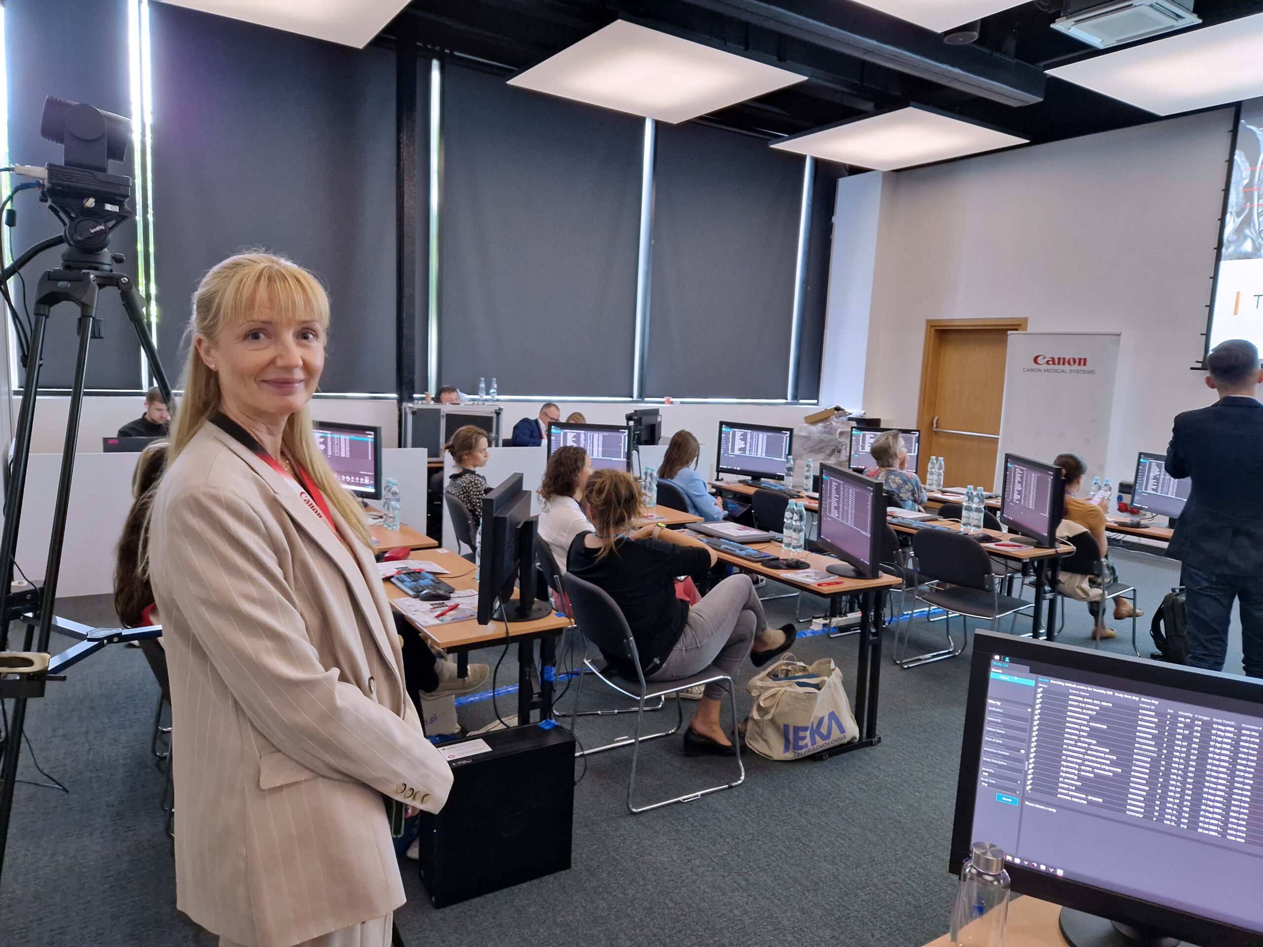
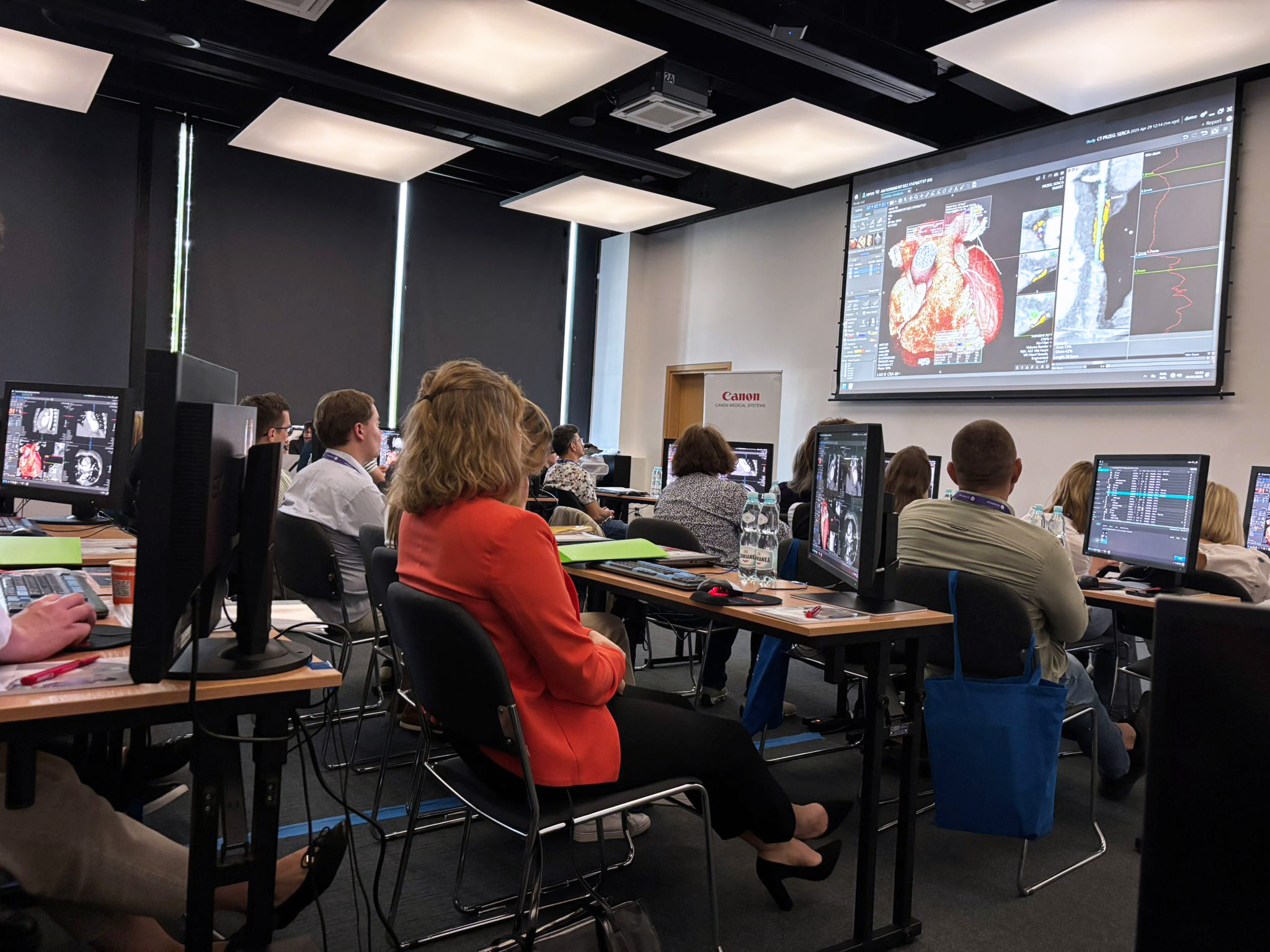
Za nami kolejna edycja Forum Elektroradiologów i Pielęgniarek Elektroradiologicznych współorganizowana przez firmę TMS (dystrybutora Canon Medical) & Bayer. Frekwencja przerosła nasze najśmielsze oczekiwania. W tegorocznej edycji udział wzięło blisko 450 uczestników!
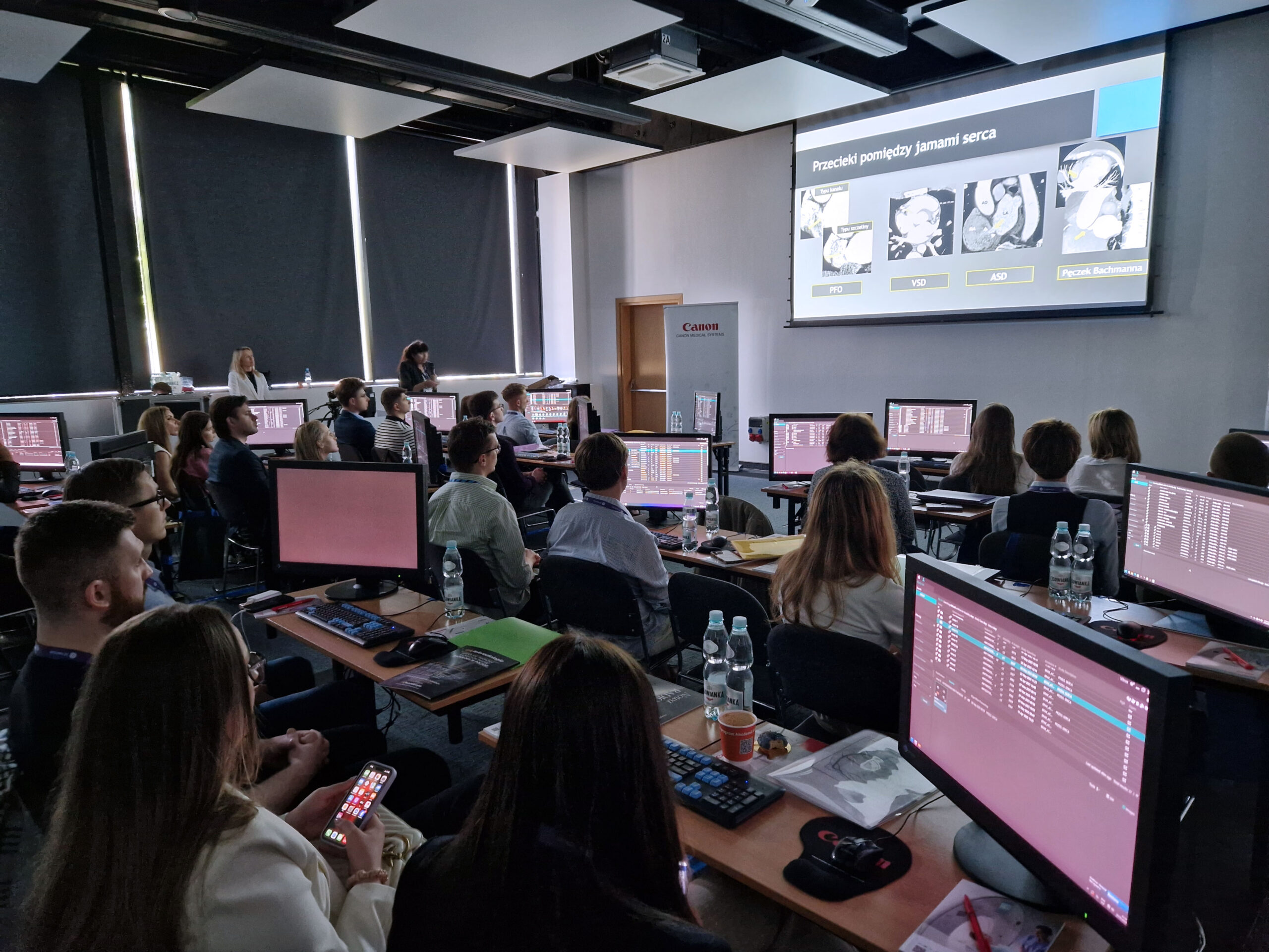
Zgodnie ze złożoną obietnicą, w ósmej edycji kontynuowaliśmy tematykę schorzeń onkologicznych, która została rozpoczęta w czasie ubiegłorocznej konferencji. W tym roku skupiliśmy się na kolejnych obszarach, zagłębiając się w takie tematy jak nowotwory płuc, przewodu pokarmowego, onkologii pediatrycznej oraz schorzeniach onkologicznych w obrębie miednicy.
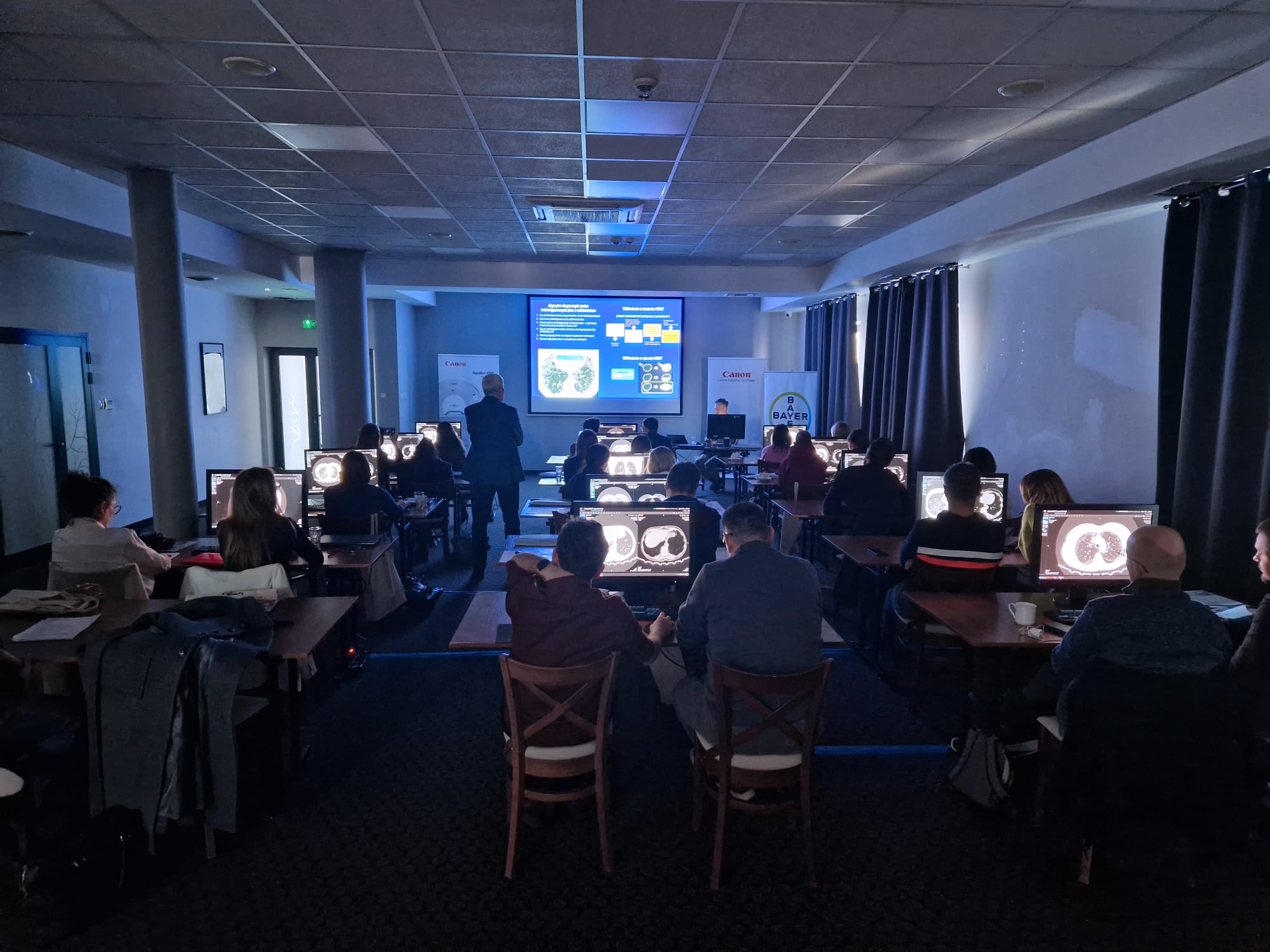
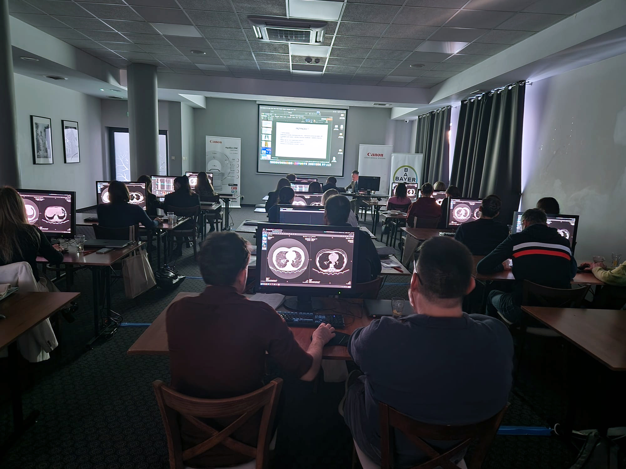
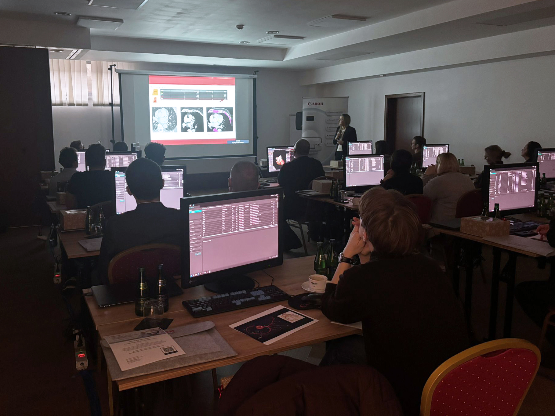
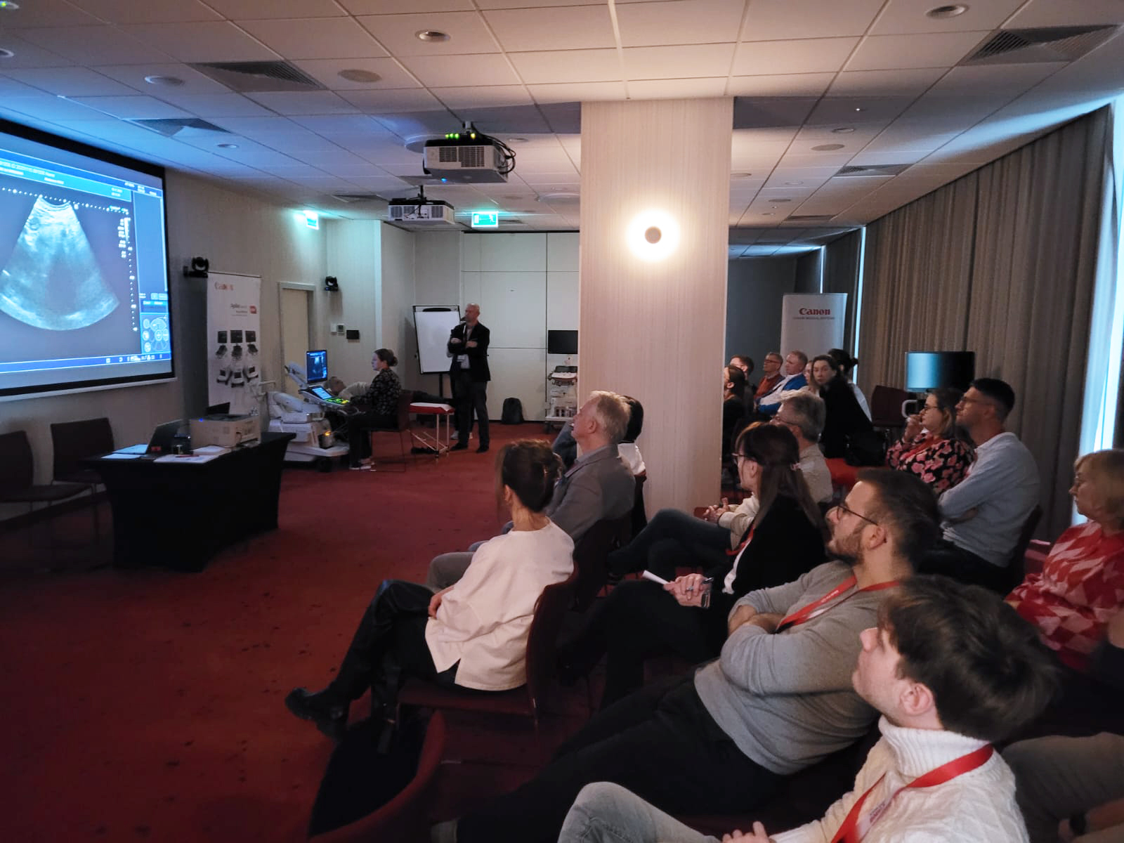
Zaproponowaliśmy Państwu blok wykładowy związany z tematyką bezpieczeństwa pacjentów onkologicznych poddawanych badaniom obrazowym, gdzie omówiliśmy zasady przygotowania do badań z podaniem środków kontrastowych, kwestię bezpieczeństwa nefrologicznego oraz postępowania w przypadku pacjentów w stanie zagrożenia życia.
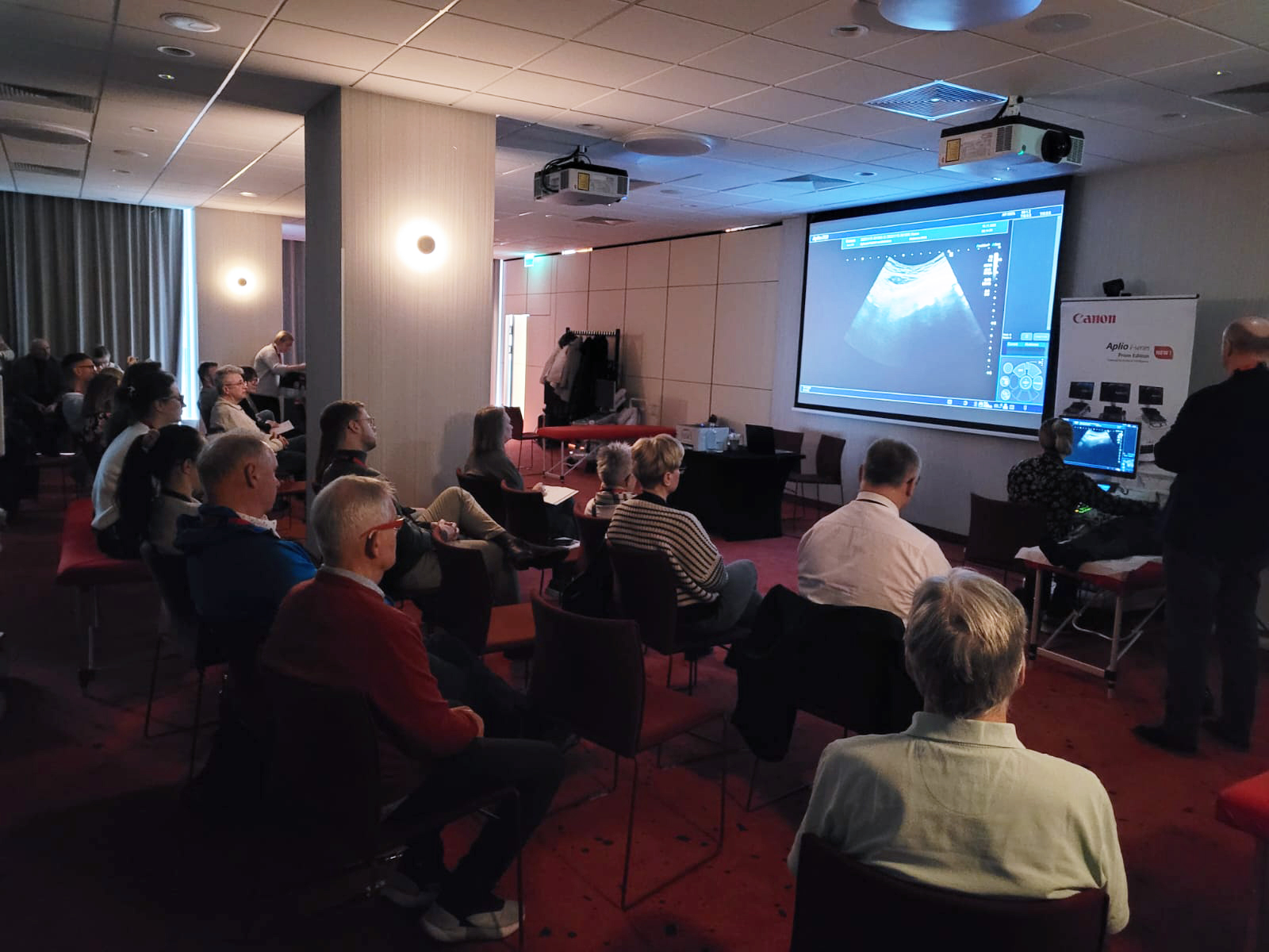
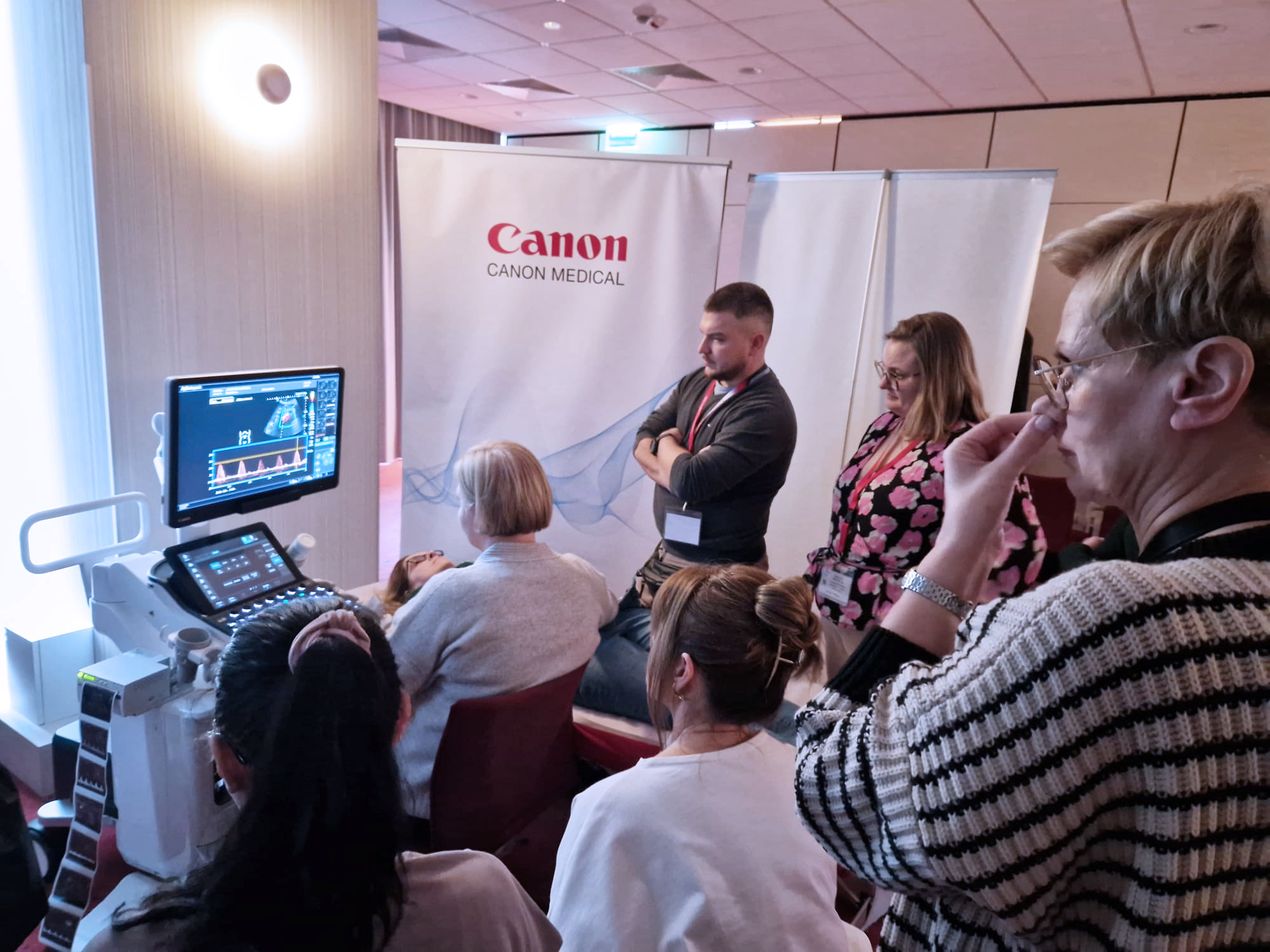
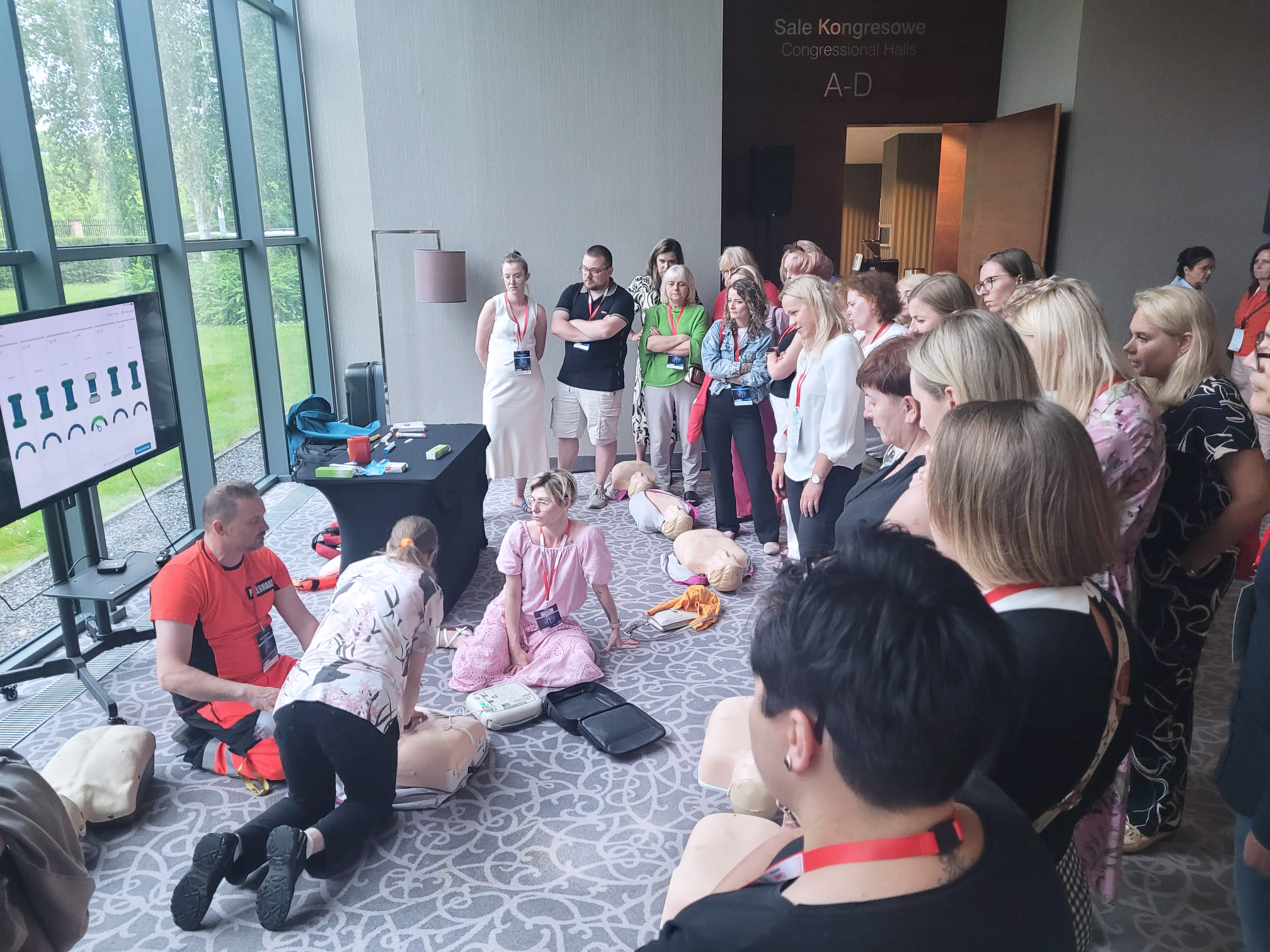
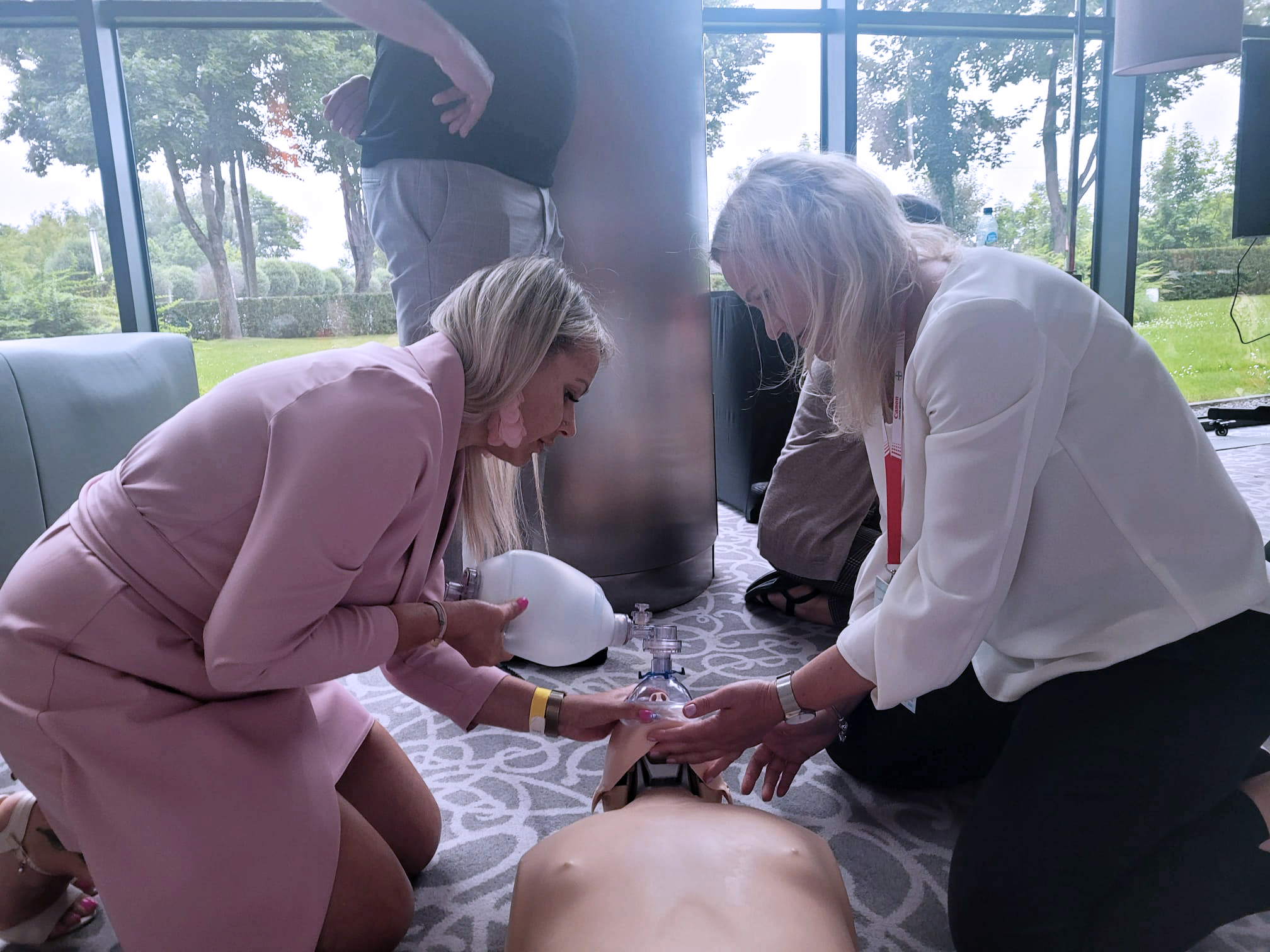
Pragnąc stworzyć przestrzeń do dyskusji oraz wspólnego poszukiwania rozwiązań przygotowaliśmy sesje wykładowe, jak i zajęcia warsztatowe.
Mamy nadzieję, że przygotowany program naukowy spełnił Państwa oczekiwania oraz zainspirował do dalszego pogłębiania tematyki.
W imieniu Rady Naukowej oraz Komitetu Organizacyjnego – dziękujemy za wspólny czas!
Przy okazji pragniemy przekazać dobrą wiadomość.
Komitet organizacyjny już wkrótce rusza z przygotowaniami do XIX Edycji Forum!
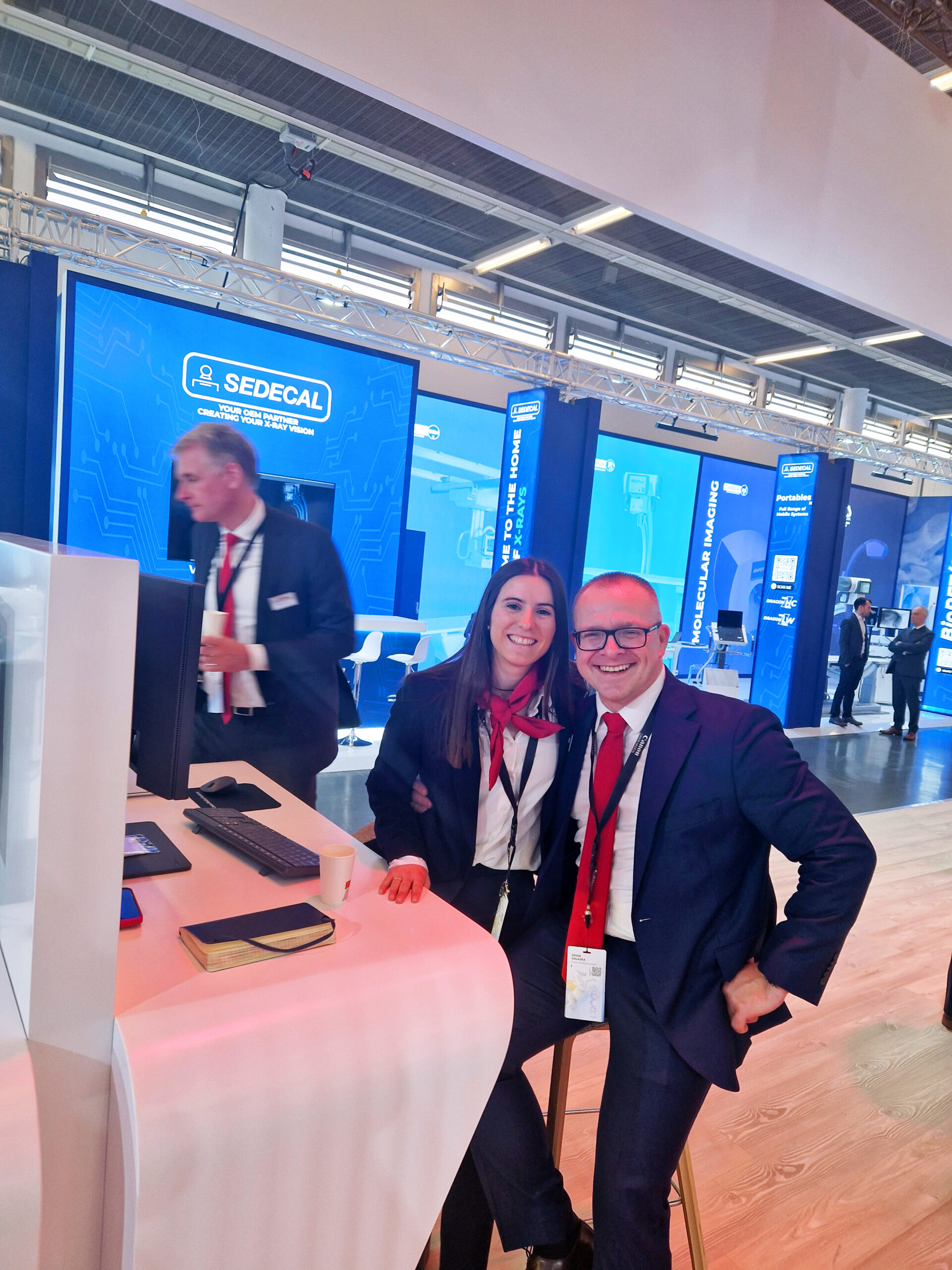
TMS Sp z o.o. czynnie uczestniczy we wspieraniu edukacji radiologicznej ?
+48 22 858 28 19
TMS Sp. z o.o.
ul. Wiertnicza 84
02-952 Warszawa,
woj. mazowieckie, Polska
NIP: 5211004948
tms@tms.com.pl
+48 22 858 28 19
+48 22 858 28 20
+48 22 858 28 12
Copyright by TMS Sp. z o.o. 2018. Wszystkie prawa zastrzeżone.
Copyright by TMS Sp. z o.o.